
Page 25 - Read Online
P. 25
Padoan et al. Vessel Plus 2021;5:41 https://dx.doi.org/10.20517/2574-1209.2021.41 Page 7 of 18
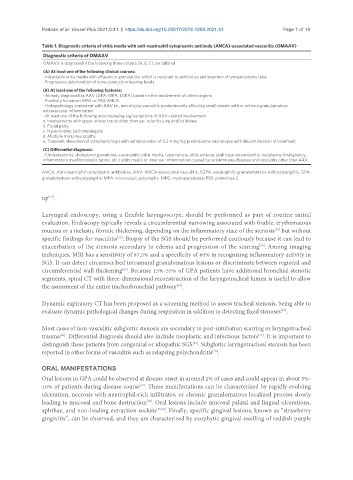
Table 1. Diagnostic criteria of otitis media with anti-neutrophil cytoplasmic antibody (ANCA)-associated vasculitis (OMAAV)
Diagnostic criteria of OMAAV
OMAVV is diagnosed if the following three criteria (A, B, C) are fulfilled
(A) At least one of the following clinical courses:
· Intractable otitis media with effusion or granulation, which is resistant to antibiotics and insertion of tympanostomy tube
· Progressive deterioration of bone conduction hearing levels
(B) At least one of the following features:
· Already diagnosed as AAV (GPA; MPA; EGPA) based on the involvement of others organs
· Positivity for serum MPO-or PR3-ANCA
· Histopathology consistent with AAV i.e., necrotizing vasculitis predominantly affecting small vessels with or without granulomatous
extravascular inflammation
· At least one of the following accompanying sig/symptoms of AAV-related involvement:
a. Involvements with upper airway tracts other than ear, scleritis lung and/or kidney
b. Facial palsy
c. Hypertrophic pachimeningytis
d. Multiple mononeuropathy
e. Transient alleviation of symptoms/sign with administration of 0.5-1 mg/Kg prednisolone and relapse with discontinuation of treatment
(C) Differential diagnosis:
· Cholesteatoma, cholesterol granuloma, eosinophilic otitis media, tuberculosis, otitis externa, skull base osteomyelitis, neoplasms (malignancy,
inflammatory myofibroblastic tumor, etc.) otitis media or inner ear inflammation caused by autoimmune diseases and vasculitis other than AAV
ANCA: Anti-neutrophil cytoplasmic antibodies; AAV: ANCA-associated vasculitis; EGPA: eosinophilic granulomatosis with polyangiitis; GPA:
granulomatosis with polyangiitis; MPA: microscopic polyangiits. MPO: myeloperoxidase; PR3: proteinase 3.
up .
[54]
Laryngeal endoscopy, using a flexible laryngoscope, should be performed as part of routine initial
evaluation. Endoscopy typically reveals a circumferential narrowing associated with friable, erythematous
[18]
mucosa or a inelastic fibrotic thickening, depending on the inflammatory state of the stenosis but without
specific findings for vasculitis . Biopsy of the SGS should be performed cautiously because it can lead to
[53]
exacerbation of the stenosis secondary to edema and progression of the scarring . Among imaging
[58]
techniques, MRI has a sensitivity of 87.5% and a specificity of 60% in recognizing inflammatory activity in
SGS. It can detect circumscribed intramural granulomatous lesions or discriminate between regional and
circumferential wall thickening . Because 15%-55% of GPA patients have additional bronchial stenotic
[43]
segments, spiral CT with three-dimensional reconstruction of the laryngotracheal lumen is useful to allow
the assessment of the entire tracheobronchial pathway .
[50]
Dynamic expiratory CT has been proposed as a screening method to assess tracheal stenosis, being able to
[47]
evaluate dynamic pathological changes during respiration in addition to detecting fixed stenoses .
Most cases of non-vasculitic subglottic stenosis are secondary to post-intubation scarring or laryngotracheal
[43]
[51]
trauma . Differential diagnosis should also include neoplastic and infectious factors . It is important to
distinguish these patients from congenital or idiopathic SGS . Subglottic laryngotracheal stenosis has been
[48]
reported in other forms of vasculitis such as relapsing polychondritis .
[46]
ORAL MANIFESTATIONS
Oral lesions in GPA could be observed at disease onset in around 2% of cases and could appear in about 5%-
[14]
10% of patients during disease course . These manifestations can be characterized by rapidly evolving
ulceration, necrosis with neutrophil-rich infiltrates, or chronic granulomatous localized process slowly
leading to mucosal and bone destruction . Oral lesions include mucosal palatal and lingual ulcerations,
[59]
aphthae, and non-healing extraction sockets [59,60] . Finally, specific gingival lesions, known as “strawberry
gingivitis”, can be observed, and they are characterized by exophytic gingival swelling of reddish purple