
Page 23 - Read Online
P. 23
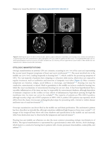
Padoan et al. Vessel Plus 2021;5:41  https://dx.doi.org/10.20517/2574-1209.2021.41  Page 5 of 18


                Figure 3. Radiological aspect of chronic rhinosinusitis, anterior septal perforation (asterisks), and maxillary sinus osteitis (arrowhead) in
                a patient diagnosed with granulomatosis with polyangiitis [CT scan, axial (A) and frontal (B) views]. T2-weighted MRI axial scan (C)
                and fluid attenuated inversion recovery (FLAIR) coronal scan (D) showing diffuse hyperintense opacification of the middle ear and
                mastoid cells in bilateral mastoiditis (asterisks).


               OTOLOGIC MANIFESTATIONS
               Otologic manifestations in systemic GPA are common, occurring in 20%-70% of the cases and representing
               the second most frequent symptoms of head and neck involvement [7,24,31] . The most involved site is the
               middle ear (23%-70%), leading frequently to hearing loss [21,32] , which could be the presenting symptom of
               GPA. The vast majority of patients have relapsing or refractory otitis media, which does not respond to
               regular treatment, such as antibiotics and insertion of tympanic vent tubes [Figure 3]. Other common
               symptoms are tinnitus, otalgia, aural fullness or discharge, and dizziness [21,33] . Hearing loss may be
               conductive, sensorineural, or mixed. Fluid or granulation in the middle ear induces conductive hearing loss,
               while the exact mechanisms of sensorineural hearing loss are not clear. It has been hypothesized that a
               vasculitic inflammation of the inner ear may be responsible for sensorineural deafness, although deposition
               of immune complexes in the cochlea or toxic effects of inflammatory byproducts passing through the
               membrane into the inner ear cannot be excluded . The majority of patients are PR3-ANCA positive,
                                                           [34]
               although recently a high rate of ENT symptoms in MPO-ANCA-positive GPA patients was reported, which
               differs from the classic microscopic polyangiitis phenotype for presenting with a limited or milder disease
               and lower rate of renal involvement [35,36] .

               Otoscopy examination can detect fluid in the middle ear and drum perforation. The audiometric pattern
               has been described as typically flat, although sometimes additional high frequency losses may coexist . CT
                                                                                                    [37]
               images of the temporal bone show soft tissue shadows and opacification in the middle ear and mastoid,
                                                                                 [21]
               while bone destruction may be observed in the tympanum and mastoid sinuses .

               Hearing loss and middle ear effusions are also the most common presenting otologic manifestations of
               EGPA. The typical manifestation is represented by a granulomatous otitis with chronic, thick discharge,
               which leads to a conductive hearing loss in patients with chronic paranasal abnormalities, eosinophilia, and
               asthma [38,39] .