


Page 57 - Read Online
P. 57
Page 4 of 10                                                   Scotti et al. Vessel Plus 2021;5:6  I  http://dx.doi.org/10.20517/2574-1209.2020.68


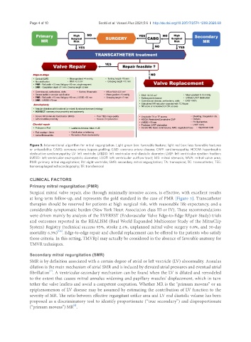
               Figure 3. Interventional algorithm for mitral regurgitation. Light green box: favorable feature; light red box: less favorable features
               or unfeasibility; CABG: coronary artery bypass grafting; CAD: coronary artery disease; CMP: cardiomyopathy; HOCM: hypertrophic
               obstructive cardiomyopathy; LV: left ventricle; LVEDD: left ventricular end-diastolic diameter; LVEF: left ventricular ejection fraction;
               LVESD: left ventricular end-systolic diameter; LVOT: left ventricular outflow tract; MS: mitral stenosis; MVA: mitral valve area;
               PMR: primary mitral regurgitation; RV: right ventricle; SMR: secondary mitral regurgitation; TA: transapical; TC: transcatheter; TEE:
               transesophageal echocardiography; TF: transfemoral


               CLINICAL FACTORS
               Primary mitral regurgitation (PMR)
               Surgical mitral valve repair, also through minimally invasive access, is effective, with excellent results
               at long-term follow-up, and represents the gold standard in the case of PMR [Figure 3]. Transcatheter
               therapies should be reserved for patients at high surgical risk, with reasonable life expectancy, and a
               considerable symptomatic burden (New York Heart Association class III or IV). These recommendations
               were driven mainly by analysis of the EVEREST (Endovascular Valve Edge-to-Edge REpair Study) trials
               and outcomes reported in the REALISM (Real World Expanded Multicenter Study of the MitraClip
               System) Registry (technical success 95%, stroke 2.4%, unplanned mitral valve surgery 0.8%, and 30-day
                            [5,6]
               mortality 6.3%) . Edge-to-edge repair and chordal replacement can be offered to the patients who satisfy
               those criteria. In this setting, TMVRpl may actually be considered in the absence of favorable anatomy for
               TMVR techniques.

               Secondary mitral regurgitation (SMR)
               SMR is by definition associated with a certain degree of atrial or left ventricle (LV) abnormality. Annulus
               dilation is the main mechanism of atrial SMR and is induced by elevated atrial pressures and eventual atrial
                        [7]
               fibrillation . A ventricular-secondary mechanism can be found when the LV is dilated and remodeled
               to the extent that causes mitral annulus widening and papillary muscles’ displacement, which in turn
               tether the valve leaflets and avoid a competent coaptation. Whether MR is the “primum movens” or an
               epiphenomenon of LV disease may be assumed by estimating the contribution of LV function to the
               severity of MR. The ratio between effective regurgitant orifice area and LV end diastolic volume has been
               proposed as a discriminatory tool to identify proportionate (“true secondary”) and disproportionate
                                    [8]
               (“primum movens”) MR .

