
Page 35 - Read Online
P. 35
Antwi-Adjei et al. Vessel Plus 2021;5:35 https://dx.doi.org/10.20517/2574-1209.2021.48 Page 7 of 10
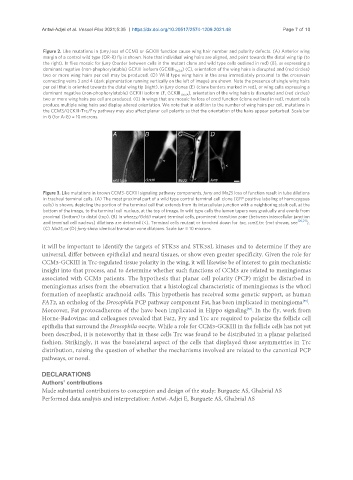
Figure 2. Like mutations in furry, loss of CCM3 or GCKIII function cause wing hair number and polarity defects. (A) Anterior wing
margin of a control wild type (OR-R) fly is shown. Note that individual wing hairs are aligned, and point towards the distal wing tip (to
the right). In flies mosaic for furry (border between cells in the mutant clone and wild type cells outlined in red) (B), or expressing a
dominant negative (non-phophorylatable) GCKIII isoform (GCKIII T167A ) (C), orientation of the wing hairs is disrupted and (red circles)
two or more wing hairs per cell may be produced. (D) Wild type wing hairs in the area immediately proximal to the crossvein
connecting veins 3 and 4 (dark pigmentation running vertically on the left of image) are shown. Note the presence of single wing hairs
per cell that is oriented towards the distal wing tip (right). In furry clones (E) (clone borders marked in red), or wing cells expressing a
dominant negative (non-phophorylatable) GCKIII isoform (F, GCKIII T167A ), orientation of the wing hairs is disrupted and (red circles)
two or more wing hairs per cell are produced. (G) In wings that are mosaic for loss of ccm3 function (clone outlined in red), mutant cells
produce multiple wing hairs and display altered orientation. We note that in addition to the number of wing hairs per cell, mutations in
the CCM3/GCKIII-Trc/Fry pathway may also affect planar cell polarity so that the orientation of the hairs appear perturbed. Scale bar
in G (for A-G) = 10 microns.
Figure 3. Like mutations in known CCM3-GCKIII signaling pathway components, furry and Mo25 loss of function result in tube dilations
in tracheal terminal cells. (A) The most proximal part of a wild type control terminal cell clone (GFP positive labeling of homozygous
cells) is shown, depicting the portion of the terminal cell that extends from its intercellular junction with a neighboring stalk cell, at the
bottom of the image, to the terminal cell nucleus, at the top of image. In wild type cells the lumen tapers very gradually and evenly from
proximal (bottom) to distal (top). (B) In wheezy/GckIII mutant terminal cells, prominent transition zone (between intercellular junction
[20,21]
and terminal cell nucleus) dilations are detected (<). Terminal cells mutant or knocked down for tao, ccm3, trc (not shown, see ),
(C) Mo25, or (D) furry show identical transition zone dilations. Scale bar = 10 microns.
it will be important to identify the targets of STK38 and STK38L kinases and to determine if they are
universal, differ between epithelial and neural tissues, or show even greater specificity. Given the role for
CCM3-GCKIII in Trc-regulated tissue polarity in the wing, it will likewise be of interest to gain mechanistic
insight into that process, and to determine whether such functions of CCM3 are related to meningiomas
associated with CCM3 patients. The hypothesis that planar cell polarity (PCP) might be disturbed in
meningiomas arises from the observation that a histological characteristic of meningiomas is the whorl
formation of neoplastic arachnoid cells. This hypothesis has received some genetic support, as human
FAT2, an ortholog of the Drosophila PCP pathway component Fat, has been implicated in meningioma .
[55]
Moreover, Fat protocadherens of the have been implicated in Hippo signaling . In the fly, work from
[56]
Horne-Badovinac and colleagues revealed that Fat2, Fry and Trc are required to polarize the follicle cell
epithelia that surround the Drosophila oocyte. While a role for CCM3-GCKIII in the follicle cells has not yet
been described, it is noteworthy that in these cells Trc was found to be distributed in a planar polarized
fashion. Strikingly, it was the basolateral aspect of the cells that displayed these asymmetries in Trc
distribution, raising the question of whether the mechanisms involved are related to the canonical PCP
pathways, or novel.
DECLARATIONS
Authors’ contributions
Made substantial contributions to conception and design of the study: Burguete AS, Ghabrial AS
Performed data analysis and interpretation: Antwi-Adjei E, Burguete AS, Ghabrial AS