


Page 120 - Read Online
P. 120
Page 10 of 14                Edwards et al. Vessel Plus 2021;5:42  https://dx.doi.org/10.20517/2574-1209.2021.18


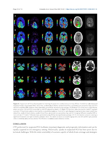
                Figure 4. A spectrum of PCS perfusion patterns involving the posterior cerebral artery territory (PCA). From left to right displayed
                imaging includes: mean transit time, delay time, cerebral blood volume, cerebral blood flow, automated core-penumbra map and 24 h
                diffusion weighted MRI. Perfusion maps generated using MiStar neuroperfusion package. (A) Bilateral PCA stroke due to distal basilar
                artery occlusion. (B) Left PCA stroke due to a left P1 segment occlusion. (C) Right PCA territory infarction due to a right P1 segment
                occlusion. PCS perfusion patterns involving the cerebellar hemispheres. From left to right displayed imaging includes: mean transit time,
                delay time, cerebral blood volume, cerebral blood flow, automated core-penumbra map and 24 h imaging (diffusion weighted MRI or
                non-contrast CT brain). Perfusion maps generated using MiStar neuroperfusion package. (D) A mid basilar occlusion causing a
                significant brainstem and right cerebellar perfusion lesion. No visible occlusion on vessel imaging with a (E) left and (F) right posterior
                inferior cerebellar artery territory stroke. PCS: Posterior circulation acute ischaemic strokes.

               CONCLUSION
               CTP performed for suspected PCS facilitates important diagnostic and prognostic information and can be
               rapidly acquired in the emergency setting. Historically, uptake in suspected PCS has been poor due to
               technical challenges. With the wider availability of scanners capable of whole brain coverage and strategies

