
Page 66 - Read Online
P. 66
Sabe et al. Vessel Plus 2024;8:2 https://dx.doi.org/10.20517/2574-1209.2023.95 Page 5 of 12
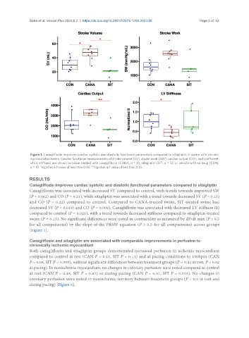
Figure 1. Canagliflozin improves cardiac systolic and diastolic functional parameters compared to sitagliptin in swine with chronic
myocardial ischemia. Cardiac functional measurements of stroke volume (SV), stroke work (SW), cardiac output (CO), and coefficient
of LV stiffness are shown in swine treated with canagliflozin (CANA, n = 8), sitagliptin (SIT, n = 5), or vehicle with no drug (CON,
n = 8). *signifies a P value of less than 0.05. **signifies a P value of less than 0.01.
RESULTS
Canagliflozin improves cardiac systolic and diastolic functional parameters compared to sitagliptin
Canagliflozin was associated with increased SV compared to control, with trends towards improved SW
(P = 0.062) and CO (P = 0.21), while sitagliptin was associated with a trend towards decreased SV (P = 0.13)
and CO (P = 0.22) compared to control. Compared to CANA-treated swine, SIT-treated swine had
decreased SV (P = 0.019) and CO (P = 0.009). Canagliflozin was associated with decreased LV stiffness (ß)
compared to control (P = 0.021), with a trend towards decreased stiffness compared to sitagliptin-treated
swine (P = 0.13). No significant differences were noted in contractility as measured by dP/dt max (P > 0.5
for all comparisons) by the slope of the PRSW equation (P > 0.5 for all comparisons) across groups
[Figure 1].
Canagliflozin and sitagliptin are associated with comparable improvements in perfusion to
chronically ischemic myocardium
Both canagliflozin and sitagliptin groups demonstrated increased perfusion in ischemic myocardium
compared to control at rest (CAN P = 0.11, SIT P = 0.11) and at pacing conditions to 150bpm (CAN
P = 0.08, SIT P = 0.005), without significant differences between treatment groups (P = 0.44 at rest, P = 0.62
at pacing). In nonischemic myocardium, no changes in coronary perfusion were noted compared to control
at rest (CAN P = 0.48, SIT P = 0.87) or during pacing (CAN P = 0.51, SIT P = 0.513). No changes in
coronary perfusion were noted in nonischemic territory between treatment groups (P > 0.5 at rest and
during pacing) [Figure 2].