
Page 194 - Read Online
P. 194
Page 6 of 16 Harik et al. Vessel Plus 2023;7:30 https://dx.doi.org/10.20517/2574-1209.2023.124
2
stented group (5.12 mm ; P = 0.04); however, at angiographic follow-up at one year, there was no difference
[39]
between groups in SVG failure (30.0% vs. 28.2%, P = 0.55) . VEST IV, which reported a 4.5-year follow-up,
similarly found no difference in SVG failure rate between stented grafts and those not stented (30.0% vs.
[40]
23.0%; P = 0.42) . VEST II studied stented SVGs grafted to the right coronary territory and found that at a
three-to-six-month follow-up, there was no difference in SVG patency between stented grafts and those not
[41]
stented (86.2% vs. 88.8%) . VEST III included 184 patients randomized to receive one stented SVG and
found no difference in graft patency between stented grafts and those not stented at two-year follow-up
[42]
(78.3% vs. 88.8%; P = 0.43) .
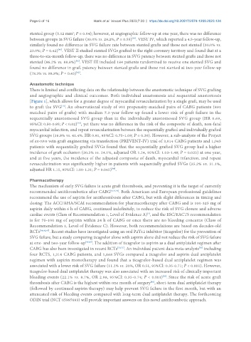
Anastomotic technique
There is limited and conflicting data on the relationship between the anastomotic technique of SVG grafting
and angiographic and clinical outcomes. Both individual anastomosis and sequential anastomosis
[Figure 1], which allows for a greater degree of myocardial revascularization by a single graft, may be used
[10]
to graft the SVG . An observational study of 901 propensity-matched pairs of CABG patients (891
matched pairs of grafts) with median 7.3-year follow-up found a lower risk of graft failure in the
sequentially anastomosed SVG group than in the individually anastomosed SVG group (HR 0.69,
95%CI: 0.50-0.95; P < 0.02) , yet there was no difference in the risk of the composite of death, non-fatal
[43]
myocardial infarction, and repeat revascularization between the sequentially grafted and individually grafted
SVG groups (36.8% vs. 41.4%. HR 0.91, 95%CI: 0.75-1.09; P = 0.30). However, a sub-analysis of the Project
of ex-vivo vein graft engineering via transfection (PREVENT-IV) trial of 3,014 CABG patients and 1,045
patients with sequentially grafted SVGs found that the sequentially grafted SVG group had a higher
incidence of graft occlusion (30.5% vs. 24.1%, adjusted OR 1.24, 95%CI: 1.03-1.48; P = 0.025) at one year,
and at five years, the incidence of the adjusted composite of death, myocardial infarction, and repeat
revascularization was significantly higher in patients with sequentially grafted SVGs (35.2% vs. 31.1%,
adjusted HR 1.15, 95%CI: 1.00-1.31; P = 0.045) .
[44]
Pharmacotherapy
The mechanism of early SVG failure is acute graft thrombosis, and preventing it is the target of currently
recommended antithrombotics after CABG [9,34,45] . Both American and European professional guidelines
recommend the use of aspirin for antithrombosis after CABG, but with slight differences in timing and
dosing: The ACC/AHA/SCAI recommendation for pharmacotherapy after CABG and is 100-325 mg of
aspirin daily within 6 h of CABG, continued indefinitely, to reduce the risk of SVG closure and adverse
cardiac events (Class of Recommendation 1, Level of Evidence A) , and the ESC/EACTS recommendation
[3]
is for 75-100 mg of aspirin within 24 h of CABG or once there are no bleeding concerns (Class of
Recommendation 1, Level of Evidence C). However, both recommendations are based on decades-old
RCTs [34,46,47] . Recent studies have investigated using an oral P2Y12 inhibitor (ticagrelor) for the prevention of
SVG failure, but a study comparing ticagrelor alone with aspirin alone did not reduce the risk of SVG failure
at one- and two-year follow-up [48,49] . The addition of ticagrelor to aspirin as a dual antiplatelet regimen after
CABG has also been investigated in recent RCTs [50,51] . An individual patient data meta-analysis including
[50]
four RCTS, 1,316 CABG patients, and 1,668 SVGs compared a ticagrelor and aspirin dual antiplatelet
regimen with aspirin monotherapy and found that a ticagrelor-based dual antiplatelet regimen was
associated with a lower risk of SVG failure (11.2% vs. 20%, OR 0.51, 95%CI: 0.35-0.71; P < 0.001). However,
ticagrelor-based dual antiplatelet therapy was also associated with an increased risk of clinically important
[50]
bleeding events (22.1% vs. 8.7%, OR 2.98, 95%CI: 0.35-0.74; P < 0.001) . Since the risk of acute graft
thrombosis after CABG is the highest within one month of surgery , short-term dual antiplatelet therapy
[45]
(followed by continued aspirin therapy) may help prevent SVG failure in the first month, but with an
attenuated risk of bleeding events compared with long-term dual antiplatelet therapy. The forthcoming
ODIN trial (NCT 05997693) will provide important answers on this novel antithrombotic approach.