Page 172 - Read Online
P. 172

Bradshaw et al. Vessel Plus 2023;7:35   https://dx.doi.org/10.20517/2574-1209.2023.121                                                                                  Page 7 of 21


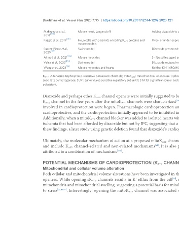
                              Makepeace et al.,      Mouse heart, Langendorff                         Adding diazoxide to cardioplegia provided improved recovery after ischemia
                                  [134]
                              2018
                                             [45]
                              Paggio et al., 2019    HeLa cells with plasmids encoding K   proteins and   Over- or under-expression of mitoK   is detrimental. Cardioprotection by diazoxide is lost when mitoK   is suppressed
                                                                                    ATP                                             ATP                                                      ATP
                                                     mouse models
                              Suarez-Pierre et al.,   Swine model                                     Diazoxide preserved systolic and diastolic ventricular function after ischemia in a large animal model
                                   [135]
                              2020
                                              [126]
                              Ahmad et al., 2021     Mouse myocytes                                   S-nitrosating agent and diazoxide are cardioprotective individually, but the beneficial effect was lost when they were combined
                                             [136]
                              Velez et al., 2022     Swine model                                      Diazoxide reduced myocardial stunning and facilitated separation from cardiopulmonary bypass
                                             [59]
                              Wang et al., 2023      Mouse myocytes and hearts                        Neither Kir1.1 (ROMK) nor SUR1 were involved in the mechanism of cardioprotection by diazoxide

                              K  : Adenosine triphosphate-sensitive potassium channels; mitoK  : mitochondrial adenosine triphosphate-sensitive potassium channels; IPC: ischemic preconditioning; 5-HD: 5-hydroxydecanoate; SDH:
                               ATP                                                       ATP
                              succinate dehydrogenase; SUR1: sulfonylurea sensitive regulatory subunit 1; STAT3: signal transducer and activator of transcription 3; SUR 1: sulfonylurea sensitive regulatory subunit 1; ROMK: renal outer medullary
                              potassium.


                              Diazoxide and perhaps other K  channel openers were initially suggested to be cardioprotective via a mechanism involving a mitoK  channel rather than a s
                                                                 ATP
                                                                                                                                                                                     ATP
                              K  channel in the few years after the mitoK  channels were characterized                [26,33] . Efforts to define the role of both sarcolemmal and mitochondrial channels
                                                                                  ATP
                               ATP
                              involved in cardioprotection were begun. Pharmacologic cardioprotection and non-pharmacologic cardioprotection using IPC were compared: both were
                              cardioprotective, and the cardioprotection initially appeared to be inhibited in the presence of a selective mitoK  channel blocker, 5-hydroxydecanoate .
                                                                                                                                                                                                              [101]
                                                                                                                                                               ATP
                              Additionally, when a mitoK  channel blocker was added to isolated hearts with either diazoxide or IPC, this abolished the improvement in contractility after
                                                             ATP
                              ischemia that had been afforded by diazoxide but not by IPC, suggesting that a mitoK  was critical for diazoxide’s mechanism but not for IPC . In line with
                                                                                                                                                                                                 [86]
                                                                                                                               ATP
                              these findings, a later study using genetic deletion found that diazoxide’s cardioprotection was not due to action at a sK  channel .
                                                                                                                                                                                 [41]
                                                                                                                                                                     ATP
                              Ultimately, the molecular mechanism of action at a proposed mitoK  channel remains unknown. Potential mechanisms largely focus on the mitochondria
                                                                                                             ATP
                              and include K  channel-related and non-related mechanisms . It is also possible that the effects of diazoxide and other K  channel openers can be
                                                                                                        [42]
                                                                                                                                                                                  ATP
                                              ATP
                              attributed to a combination of mechanisms .
                                                                               [102]
                              POTENTIAL MECHANISMS OF CARDIOPROTECTION (K  CHANNEL AND CHANNEL-INDEPENDENT)
                                                                                                          ATP
                              Mitochondrial and cellular volume alteration
                              Both cellular and mitochondrial volume alterations have been investigated in the search for mechanisms of cardioprotection relating to K  channels and their
                                                                                                                                                                                          ATP
                                                                                                                    [30]
                                                                                                                                                                           +
                              openers. While opening sK  channels results in K  efflux from the cell , opening of a mitoK  channel results in K  influx from the cytosol into the
                                                                                           +
                                                                                                                                               ATP
                                                              ATP
                              mitochondria and mitochondrial swelling, suggesting a potential basis for mitoK -dependent changes in mitochondrial and cellular volume alteration during
                                                                                                                         ATP
                              to stress [32,46,67] . Interestingly, opening the mitoK  channel was associated with changes in mitochondrial matrix volume, calcium concentration, and
                                                                                       ATP
   167   168   169   170   171   172   173   174   175   176   177