


Page 43 - Read Online
P. 43
Trahanas et al. Vessel Plus 2022;6:49  https://dx.doi.org/10.20517/2574-1209.2021.125  Page 3 of 16


                                                                          [3]
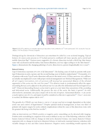
                Figure 1. SVS/STS guidelines on anatomic reporting of dissections from Lombardi et al.  (with permission). SVS: Society for Vascular
                Surgery. STS: The Society of Thoracic Surgeons.

               Distinguishing the chronicity of dissection can sometimes be aided by cross-sectional imaging. Typical
               features of a more acute process on CT include periaortic confluent soft tissue opacity, and a curved and
               mobile dissection flap . Features more suggestive of a chronic dissection include a thick flap, false lumen
                                  [5]
               outer wall calcification and thrombus, false lumen dilatation, and tear edges curling in to the false lumen .
                                                                                                        [5]
               Figure 2 demonstrates the changing pathology of aortic dissection in a patient longitudinally over 4 years .
                                                                                                       [6]
               Natural history
               Type B dissections represent 25%-40% of all dissections . The leading cause of death in patients with acute
                                                              [4]
                                                                                         [7]
               type B dissection is aortic rupture, and the second leading cause of death is malperfusion . Fortunately, 91%
               of patients with acute Type B aortic dissection will survive the initial event. Of these survivors, 66% will have
               an initially uncomplicated course with proper medical management (see Medical Management). While not
               all will require intervention, 60% of survivors will develop late aneurysmal dilation of the affected
               segment(s) of the aorta over the next five years . This dilation tends to predominantly affect the upper
                                                         [8]
               descending thoracic aorta adjacent to the primary tear but can affect the abdominal and visceral segments as
               well . Dissected descending thoracic aortas tend to grow at a rate faster than aneurysms of the ascending
                   [7]
                                                                                                [9]
               and abdominal aorta. Additionally, the greater the size of the aorta, the faster it grows . As with
               atherosclerotic aneurysms, size plays a major role in the risk of rupture. Aneurysmal dilation > 6 cm carries
               a yearly rupture rate of 6.9%, and repair should be considered as this size is approached . Once 7 cm in size
                                                                                        [10]
                                                  [9]
               is reached, the lifetime rupture risk is 43% .
               The growth of a CTBAD can vary from 0.1 cm to 0.7 cm per year but is strongly dependent on the initial
               aortic size and control of hypertension . Despite optimal medical management, at least one-third of
                                                  [9]
               patients will require surgery for aortic-related complications within five years of the dissection event, with
               the greatest risk being in the first few months .
                                                     [2]

               After an intervention on CTBAD, positive or negative aortic remodeling can be detected with imaging.
               Positive aortic remodeling in a segment of the aorta is defined as any one of the following: reduction in false
               lumen diameter/volume with no change in total aortic diameter/volume; true lumen diameter/volume
               expansion with no change in total aortic diameter/volume; or total aortic maximal diameter reduction with
               variable changes in true and false lumen diameters. Negative aortic remodeling would represent the

