


Page 56 - Read Online
P. 56
Page 4 of 10                 Dall’Ara et al. Vessel Plus 2023;7:26  https://dx.doi.org/10.20517/2574-1209.2023.107

               Table 1. Baseline characteristics
                                    Overall   Fem                                              1     2
                Variable                                Rad_Stand 114 (51.1)  Rad_SGC 37 (16.6)  P  P
                                    n = 223   72 (32.2)
                Age, years          74.8 ± 9.1  74.8 ± 9.1  74.0 ± 9.2      74.7 ± 8.4        0.259  0.700
                Male gender         162 (72.6)  47 (65.3)  92 (80.7)        23 (62.2)         0.021  0.021
                Hypertension        193 (86.5)  62 (86.1)  99 (86.8)        32 (86.5)         0.990  1.000
                Hypercholesterolemia  166 (74.4)  52 (72.2)  88 (77.8)      26 (70.3)         0.613  0.395
                Current smoker      80 (35.9)  28 (38.9)  37 (32.5)         15 (40.5)         0.545  0.369
                         2
                BMI > 30 Kg/m       51 (22.9)  17 (23.6)  29 (25.4)         5 (13.5)          0.319  0.131
                Diabetes mellitus   97 (43.5)  32 (44.4)  52 (45.6)         13 (35.1)         0.525  0.263
                Dialyses            13 (5.8)  7 (9.7)   6 (5.3)             0 (0.0)           0.114  0.337
                PAD                 78 (35.0)  30 (41.7)  35 (30.7)         13 (35.1)         0.311  0.615
                Ejection fraction, %  50 (40-55)  50 (42-55)  50 (45-55)    50 (40-60)        0.416  0.503
                Ejection fraction < 30%  14 (6.4)  8 (11.3)  3 (2.7)        3 (8.3)           0.060  0.155
                Clinical presentation:                                                        0.101  0.051
                - ACS               99 (44.4)   29 (40.3)   58 (50.9)       12 (32.4)
                - Stable/staged     124 (55.6)  43 (59.7)  56 (49.1)        25 (67.6)
                Antithrombotic therapy:
                - Vit.K ant./DOAC    25 (11.2)   12 (16.7)   9 (7.9)        4 (10.8)          0.181   0.522
                - DAPT pretreatment  68 (30.5)  22 (30.6)  32 (28.1)        14 (37.8)         0.533  0.262

               Categorical variables are shown as number (percentage); continuous variables as median (interquartile range) or mean value (±standard
               deviation). ACS: Acute coronary syndrome; BMI: body mass index; DAPT: double antiplatelet therapy; DOAC: direct oral anticoagulant; Fem:
               femoral access; PAD: peripheral artery disease; Rad_SGC: radial sheathless guiding catheter access; Rad_Stand: radial standard access; Vit.K ant:
                             1                          2
               vitamin K antagonist; P : comparison between the three groups; P : comparison between the two radial groups.


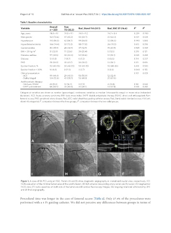
                Figure 1. A case of RA PCI using an SGC. Panels (A and B) show diagnostic angiography in cranial and caudal view, respectively; (C)
                IVUS evaluation of the minimal lumen area of the calcific lesion; (D) left anterior descending artery ostial calcific lesion; (E) longitudinal
                IVUS view; (F) radio-opacities on both side of the lumen on still cardiac fluoroscopy images; (G) ongoing rotational atherectomy; (H)
                and (I) final angiography.

               Procedural time was longer in the case of femoral access [Table 2]. Only 27.4% of the procedures were
               performed with a 6 Fr guiding catheter. We did not perceive any differences between groups in terms of

