


Page 33 - Read Online
P. 33
Andò et al. Vessel Plus 2023;7:15  https://dx.doi.org/10.20517/2574-1209.2023.26  Page 15 of 22


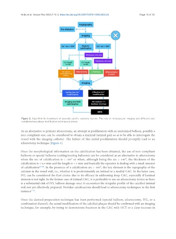
                Figure 3. Algorithm for treatment of severely calcific coronary lesions. The role of intravascular imaging and different and
                complementary plaque modification techniques is shown.


               As an alternative to primary atherectomy, an attempt at predilatation with an undersized balloon, possibly a
               non-compliant one, can be considered to obtain a minimal luminal gain so as to be able to interrogate the
               vessel with the imaging catheter. The failure of this initial predilatation should promptly lead to an
               atherectomy technique [Figure 3].

               Once the morphological information on the calcification has been obtained, the use of non-compliant
               balloons or special balloons (cutting/scoring balloons) can be considered as an alternative to atherectomy
               when the arc of calcification is < 180° or when, although being the arc > 180°, the thickness of the
               calcification is < 0.5 mm and the length is < 5 mm and basically the operator is dealing with a small amount
               of calcification [27,29] . In the presence of a calcification arc > 180°, the key element is the topography of the
               calcium in the vessel wall, i.e., whether it is predominantly an intimal or a medial CAC. In the latter case,
               IVL can be considered the first choice due to its efficacy in addressing deep CAC, especially if luminal
               stenosis is not tight. In the former case of intimal CAC, it is preferable to use an atherectomy device as there
               is a substantial risk of IVL balloon damage once it encounters the irregular profile of the calcified intimal
               wall not yet effectively prepared. Nodular calcifications should lead to atherectomy techniques in the first
               instance .
                      [115]

               Once the desired preparation technique has been performed (special balloon, atherectomy, IVL, or a
               combination thereof), the actual modification of the calcified plaque should be confirmed with an imaging
               technique, for example, by trying to demonstrate fractures in the CAC with OCT or a clear increase in

