
Page 7 - Read Online
P. 7
Brown. Rare Dis Orphan Drugs J. 2025;4:21 https://dx.doi.org/10.20517/rdodj.2025.14 Page 3 of 15
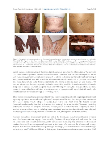
Figure 1. Examples of cutaneous neurofibromas. Moderate to severe burden of average-sized cutaneous neurofibromas on a male with
Fitzpatrick Skin Type 6 on (A) chest and abdomen, and (B) hands; (C) and (D) Moderate burden of average-sized cutaneous
neurofibromas on the chest and hands, respectively, of a woman with Fitzpatrick Skin Type 1; (E) Mild case of cutaneous neurofibromas
(arrow heads) on a background of excess skin freckling; (F) A moderate-severe case of cutaneous neurofibromas on the abdomen.
Ruler indicates approximate size of the lesions.
sample analyzed by the pathologist; therefore, clinical acumen is important for differentiation. The nerves in
CNs include both myelinated and non-myelinated axons. Compared with the surrounding skin, CNs are
well-vascularized, containing small arterioles as well as arterial and venous capillaries typically consisting of
a single endothelial cell layer with or without subendothelial smooth muscle cells or pericytes, surrounded
by a vessel basal lamina and a thickened adventitia. The intima and media layers are also enlarged. On
electron microscopy, there are corpuscle-like bodies surrounding small blood vessels or myelinated vessels,
composed of lamellar Schwann and perineurial cells with long processes, fine collagen fibers, and basal
lamina. A pyramidal cell type with long tripartite processes in connection with morphologically similar cells
[7]
was identified on electron microscopy as a “covering cell” .
These tumors contain a high percentage of infiltrating tumor-supporting cells with atypical proliferative and
signaling capabilities associated with haploinsufficiency of neurofibromin from the germline mutation of
NF1. Aside from spindle-shaped Schwann-like tumor cells that form the tumor stroma
(immunohistochemically identified by Sox10 or S100 staining), there are plentiful fibroblasts (including
endoneurial fibroblast-like cells) and perineurial-like cells, as well as endothelial cells and pericytes. There is
a robust immune cell component including tumor-associated lymphocytes, dendritic cells, mast cells, and
macrophages [8-11] . Occasionally, extensive eosinophils are seen, although this is not characteristic.
Schwann-like cells do not normally proliferate within the dermis, and thus, the identification of tumor
stromal cells in a cutaneous biopsy - characterized by fusiform cells irregularly distributed within the ECM
on hematoxylin and eosin (H&E) staining or by immunohistochemical (IHC) detection of Schwann cell
markers S100 and Sox-10 - is generally accepted as diagnostic of a tumor of the Schwann cell lineage.
Beyond this classic neurofibroma histopathologic phenotype, additional more unusual morphologic
variants also exist . CNs are difficult to distinguish from cutaneous schwannomas on routine H&E
[12]