
Page 6 - Read Online
P. 6
Page 2 of 15 Brown. Rare Dis Orphan Drugs J. 2025;4:21 https://dx.doi.org/10.20517/rdodj.2025.14
such as peripheral nerve sheath tumors of the Schwann cell ontology, aberrant bone formation/
[1,2]
reabsorption, and low-grade gliomas of the brain, as well as other tumors of neural crest-derived tissues .
The protein product of NF1, neurofibromin, serves to positively regulate adenylyl cyclase, inactivate rat
sarcoma (Ras), inhibit mitosis and induce apoptosis, and inhibit cell adhesion and motility. With germline
haploinsufficiency of neurofibromin, Ras signaling escalates, leading to downstream mitogen-activated
protein kinase kinase (MEK) activation. Cutaneous neurofibromas (CNs) are benign tumors of the dermis
that arise from biallelic loss of NF1 in Schwann cell-like tumor cells and occur in 99% of NF1 patients. They
typically begin to develop in late childhood/early adolescence and have self-limited growth trajectories, but
lifetime accumulation can ultimately involve the entire integument. CNs can remain small/barely
perceptible, or they may grow to large sizes, sometimes reaching many centimeters in diameter. Typically,
each patient exhibits a characteristic number and size distribution of tumors, which can range from just a
few to tens of thousands. Although individual tumors eventually enter a state of senescence, their ultimate
size cannot be precisely predicted.
CNs are primarily associated with physical deformity that can lead to embarrassment, a tendency to cover
up the skin with bulky clothing, barriers to intimacy, and socioeconomic disparity due to fewer
opportunities for client-facing jobs. CNs can be pruritic or painful and may catch on clothing or jewelry,
resulting in bleeding, swelling, and irritation. NF1 quality of life (QOL) metrics have identified that CNs
[3-5]
play a major role in the negative impact of NF1 on mood and social interaction . The scientific
establishment has recognized the importance of understanding CN biology vis-à-vis other tumor types, and
[6]
of identifying tolerable treatments with satisfactory outcomes for patients . This review article summarizes
what is known about CNs, including appearance, tumor initiation, preclinical models, the ultrastructure of
CNs, and barriers/challenges of developing further therapies.
CNS HAVE A RANGE OF APPEARANCES
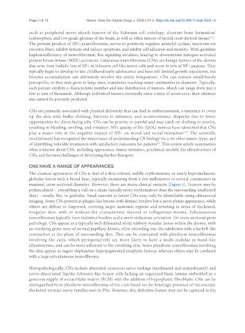
The classical appearance of CNs is that of a skin-colored, mildly erythematous, or rarely hypermelanotic
globular lesion with a broad base, typically measuring from a few millimeters to several centimeters in
maximal cross-sectional diameter. However, there are many clinical variants [Figure 1]. Tumors may be
pedunculated - resembling a ball on a chain (usually more erythematous than the surrounding unaffected
skin) - sessile, flat, or globular. Small nascent or latent CNs may only be identifiable using ultrasound
imaging. Some CNs present as plaque-like lesions with distinct borders but a more planar appearance, while
others are diffuse or dispersed, covering larger anatomic regions and resulting in areas of thickened,
irregular skin, with or without the characteristic myxoid or collagenous stroma. Subcutaneous
neurofibromas typically have indistinct borders and a more violaceous coloration. On cross-sectional gross
pathology, CNs appear as a typically well-delineated white rubbery nodular lesion within the dermis, with
an overlying grenz zone of normal papillary dermis, often extending into the subdermis with a barbell-like
contraction at the plane of surrounding skin. This can be contrasted with plexiform neurofibromas
involving the cutis, which perioperatively are more likely to have a multi-nodular or band-like
ultrastructure, and can be more adherent to the overlying skin. Some plexiform neurofibromas involving
the skin appear as rugate elephantine hyperpigmented exophytic lesions, whereas others may be confused
with a large subcutaneous neurofibroma.
Histopathologically, CNs include abnormal cutaneous nerve endings (myelinated and unmyelinated) and
nerve-dissociated bipolar Schwann-like tumor cells lacking an organized basal lamina embedded in a
generous supply of extracellular matrix (ECM) with the addition of hyperplastic fibroblasts. CNs can be
distinguished from plexiform neurofibromas of the cutis based on the histologic presence of microscopic
thickened vermian nerve bundles seen in PNs. However, this definitive feature may not be captured in the