
Page 98 - Read Online
P. 98
Page 10 of 34 West et al. Rare Dis Orphan Drugs J 2024;3:22 https://dx.doi.org/10.20517/rdodj.2023.61
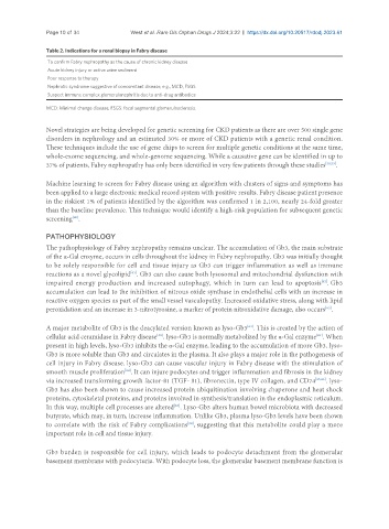
Table 2. Indications for a renal biopsy in Fabry disease
To confirm Fabry nephropathy as the cause of chronic kidney disease
Acute kidney injury or active urine sediment
Poor response to therapy
Nephrotic syndrome suggestive of concomitant disease, e.g., MCD, FSGS
Suspect immune complex glomerulonephritis due to anti-drug antibodies
MCD: Minimal change disease; FSGS: focal segmental glomerulosclerosis.
Novel strategies are being developed for genetic screening for CKD patients as there are over 500 single gene
disorders in nephrology and an estimated 30% or more of CKD patients with a genetic renal condition.
These techniques include the use of gene chips to screen for multiple genetic conditions at the same time,
whole-exome sequencing, and whole-genome sequencing. While a causative gene can be identified in up to
37% of patients, Fabry nephropathy has only been identified in very few patients through these studies [58,59] .
Machine learning to screen for Fabry disease using an algorithm with clusters of signs and symptoms has
been applied to a large electronic medical record system with positive results. Fabry disease patient presence
in the riskiest 1% of patients identified by the algorithm was confirmed 1 in 2,100, nearly 24-fold greater
than the baseline prevalence. This technique would identify a high-risk population for subsequent genetic
[60]
screening .
PATHOPHYSIOLOGY
The pathophysiology of Fabry nephropathy remains unclear. The accumulation of Gb3, the main substrate
of the α-Gal enzyme, occurs in cells throughout the kidney in Fabry nephropathy. Gb3 was initially thought
to be solely responsible for cell and tissue injury as Gb3 can trigger inflammation as well as immune
[61]
reactions as a novel glycolipid . Gb3 can also cause both lysosomal and mitochondrial dysfunction with
[62]
impaired energy production and increased autophagy, which in turn can lead to apoptosis . Gb3
accumulation can lead to the inhibition of nitrous oxide synthase in endothelial cells with an increase in
reactive oxygen species as part of the small vessel vasculopathy. Increased oxidative stress, along with lipid
peroxidation and an increase in 3-nitrotyrosine, a marker of protein nitroxidative damage, also occurs .
[63]
A major metabolite of Gb3 is the deacylated version known as lyso-Gb3 . This is created by the action of
[64]
[48]
[64]
cellular acid ceramidase in Fabry disease . lyso-Gb3 is normally metabolized by the α-Gal enzyme . When
present in high levels, lyso-Gb3 inhibits the α-Gal enzyme, leading to the accumulation of more Gb3. lyso-
Gb3 is more soluble than Gb3 and circulates in the plasma. It also plays a major role in the pathogenesis of
cell injury in Fabry disease. lyso-Gb3 can cause vascular injury in Fabry disease with the stimulation of
smooth muscle proliferation . It can injure podocytes and trigger inflammation and fibrosis in the kidney
[64]
via increased transforming growth factor-ß1 (TGF- ß1), fibronectin, type IV collagen, and CD74 [65,66] . lyso-
Gb3 has also been shown to cause increased protein ubiquitination involving chaperone and heat shock
proteins, cytoskeletal proteins, and proteins involved in synthesis/translation in the endoplasmic reticulum.
In this way, multiple cell processes are altered . Lyso-Gb3 alters human bowel microbiota with decreased
[67]
butyrate, which may, in turn, increase inflammation. Unlike Gb3, plasma lyso-Gb3 levels have been shown
to correlate with the risk of Fabry complications , suggesting that this metabolite could play a more
[68]
important role in cell and tissue injury.
Gb3 burden is responsible for cell injury, which leads to podocyte detachment from the glomerular
basement membrane with podocyturia. With podocyte loss, the glomerular basement membrane function is