
Page 842 - Read Online
P. 842
Page 4 of 12 Favre et al. Plast Aesthet Res 2020;7:71 I http://dx.doi.org/10.20517/2347-9264.2020.149
Figure 1. Injection sites for forehead rhytids into the frontalis muscle
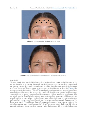
Figure 2. Injection sites for glabellar rhytids into the procerus and corrugator supercilii muscle
Lateral orbit
The main muscle of the lateral orbit is the orbicularis oculi muscle, the muscle involved in closure of the
eye and depression of the eyebrow. This muscle is divided into three parts: the orbital, the preseptal, and
the pretarsal portions. The muscle extends beyond the orbital rim and creates lateral rhytids known as
crow’s feet. Treatment of these rhytids can be done with one or three injections on either side [Figure 3]. In
a two-center randomized study by Fabi et al. , no statistically significant difference was seen in crow’s feet
[15]
treated with one injection per side vs. crow’s feet treated with three injections per side. In addition, there
was no difference in adverse events based on injection number. With the use of three injections per side,
the middle injection should be in line with the lateral canthus and the remaining should be 8 to 10 mm
away. It is critical to inject at least 1 cm lateral to the lateral orbital rim in order to avoid complications,
such as diplopia or strabismus, from diffusion into the extraocular muscles resulting in the paralysis of the
lateral rectus muscle . In addition to the crow’s feet rhytids, hypertrophy of the pretarsal portion of the
[13]
orbicularis oculi can cause what is known as the “jelly roll” appearance around the lower eyelid. When a
person is smiling, the contraction of the pretarsal portion diminishes the side of the palpebral aperture