
Page 66 - Read Online
P. 66
Liu et al. Plast Aesthet Res 2020;7:6 I http://dx.doi.org/10.20517/2347-9264.2019.62 Page 3 of 7
Figure 1. Physical examination can provide useful information on the degree of fluid retention in the edematous limb. In this patient with
left upper-limb breast cancer-related lymphedema, her limb was soft and there was no indentation after pressing on the skin. These
findings signified that the limb was fat predominate. Patients with fatty phase of lymphedema are the ideal candidates for liposuction
A B
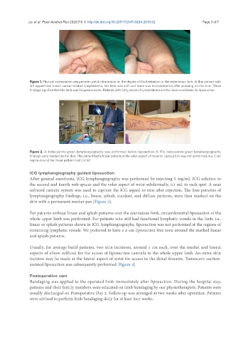
Figure 2. A: Indocyanine green-lymphangiography was performed before liposuction; B: The indocyanine green lymphangiography
findings were marked on the skin. This patient had a linear pattern at the volar aspect of forearm. Liposuction was not performed in a 2-cm
region around the linear pattern (red circle)
ICG lymphangiography guided liposuction
After general anesthesia, ICG lymphangiography was performed by injecting 5 mg/mL ICG solution to
the second and fourth web-spaces and the volar aspect of wrist subdermally, 0.1 mL to each spot. A near
infrared camera system was used to capture the ICG signal 30 min after injection. The four patterns of
lymphangiography findings, i.e., linear, splash, stardust, and diffuse patterns, were then marked on the
skin with a permanent marker pen [Figure 2].
For patients without linear and splash patterns over the edematous limb, circumferential liposuction of the
whole upper limb was performed. For patients who still had functional lymphatic vessels in the limb, i.e.,
linear or splash patterns shown in ICG lymphangiography, liposuction was not performed at the regions of
remaining lymphatic vessels. We preferred to have a 2-cm liposuction-free zone around the marked linear
and splash patterns.
Usually, for average build patients, two skin incisions, around 1 cm each, over the medial and lateral
aspects of elbow sufficed for the access of liposuction cannula to the whole upper limb. An extra skin
incision may be made at the lateral aspect of wrist for access to the distal forearm. Tumescent suction-
assisted liposuction was subsequently performed [Figure 3].
Postoperative care
Bandaging was applied to the operated limb immediately after liposuction. During the hospital stay,
patients and their family members were educated on limb bandaging by our physiotherapists. Patients were
usually discharged on Postoperative Day 2. Follow-up was arranged at two weeks after operation. Patients
were advised to perform limb bandaging daily for at least four weeks.