
Page 642 - Read Online
P. 642
Fung et al. Plast Aesthet Res 2020;7:55 I http://dx.doi.org/10.20517/2347-9264.2020.60 Page 3 of 5
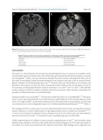
A B
Figure 3. Magnetic resonance imaging scan (axial view) reveals a T1 hypointense (A) and T2 hyperintense (B) fluid filled cystic
structure in the left upper eyelid consistent with Wolfring dacryops
Table 1. Summary table of previously reported cases of dacryops of the accessory lacrimal gland [8,10,11,15-22]
Unilateral 48 out of 49 (98%)
Bilateral 1 out of 49 (2%)
Upper eyelid 35 out of 49 (71%)
Lower eyelid 14 out of 49 (29%)
DISCUSSION
Dacryops is a closed ductal cyst arising from lacrimal gland tissue. It presents as a painless, well-
circumscribed, translucent lesion often with a bluish tint, and it may be derived from the main or accessory
[8]
lacrimal glands . These cysts are commonly classified by their location, with palpebral lobe cysts of
the main lacrimal gland (simple dacryops) being the most common and cysts of the accessory lacrimal
glands of Krause and Wolfring being quite rare [9-11] . However, there are limited data on its incidence and
prevalence. The majority of cases have been reported from Saudi Arabia, where the prevalence of cysts of
[10]
[11]
the accessory lacrimal glands has been found to be between 1 in 6,800 and 1 in 7000 . The estimates
in these reports are based on studies of patients seen in an oculoplastic clinic and may overestimate the
prevalence of these lesions due to ascertainment bias.
Dacryops usually occur unilaterally [12,13] , but they have rarely been shown to occur bilaterally [14,15] . The upper
eyelid is more commonly involved (70.8%) than the lower eyelid, likely due to the fact that there are more
[8]
ducts in the upper eyelid . In previously reported cases of dacryops of the accessory glands, the mean age
of occurrence was 30.5 years (ranging from ages 2 to 81), without male/female predominance [8,10,11,15-22] [Table 1].
The etiology of WD is unclear, and it has been hypothesized that conjunctival scarring (from previous
trauma, infection, or chronic inflammation) or IgA hypersecretion may contribute to its formation [10-12,21] .
A review of the literature by Galindo-Ferreiro estimates that up to 85% of dacryops have been linked to
[11]
previous conjunctival scarring . In our patient’s case, no predisposing cause was identified.
While congenital ptosis in children is more common, acquired ptosis is rare [2,23] and warrants a more
detailed exam and history. Eversion of the eyelid made the clinical diagnosis of WD in this case, based on
the mobile, non-tender mass located near the superior tarsal border. MRI findings can be supportive and