
Page 641 - Read Online
P. 641
Page 2 of 5 Fung et al. Plast Aesthet Res 2020;7:55 I http://dx.doi.org/10.20517/2347-9264.2020.60
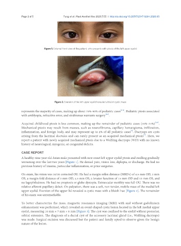
Figure 1. External front view of the patient, who presents with ptosis of the left upper eyelid
Figure 2. Eversion of the left upper eyelid revealed a bluish cystic mass
[2-4]
represents the majority of cases, making up about 79%-90% of pediatric cases . Pediatric ptosis associated
with amblyopia, refractive error, and strabismus warrants surgery .
[5,6]
[2,3]
Acquired childhood ptosis is less common, making up the remainder of pediatric cases (10%-21%) .
Mechanical ptosis may result from masses, such as neurofibroma, capillary hemangioma, infiltration,
inflammation, and foreign body, and may represent up to 2% of all pediatric cases . Dacryops are cysts
[3]
[7]
arising from the lacrimal ductules and can rarely present as an acquired mechanical ptosis . Here, we
report a patient with newly acquired mechanical ptosis due to a Wolfring dacryops (WD) with no known
history of neurological, myogenic, or congenital deficits.
CASE REPORT
A healthy nine-year old Asian male presented with new onset left upper eyelid ptosis and swelling gradually
worsening over the last two years [Figure 1]. He denied pain, vision loss, diplopia, or discharge. He had no
previous history of trauma, periocular inflammation, or prior surgeries.
On exam, his vision was 20/20 corrected OU. He had a margin reflex distance (MRD1) of 4.5 mm OD, 2 mm
OS, a margin fold distance of 3 mm OD, 4.5 mm OS, a levator function of 15 mm OD and 13 mm OS, and
no lagophthalmos. He had no proptosis or globe dystopia. Extraocular motility was full OU. There was no
relative afferent pupillary defect. On palpation, there was a soft, non-tender, mobile mass of the medial left
upper eyelid. Eversion of the upper lid revealed a cystic mass with a bluish hue [Figure 2]. The remainder
of his exam was unremarkable.
To better characterize the mass, magnetic resonance imaging (MRI) with and without gadolinium
enhancement was performed, which revealed an ovoid-shaped cystic lesion located in the left medial upper
eyelid, measuring 10 mm × 7 mm × 8 mm [Figure 3]. The cyst was confined to the eyelid without posterior,
orbital extension. The diagnosis of a ductal cyst of the accessory lacrimal gland (i.e., Wolfring dacryops)
was made. Surgical excision was discussed but the patient and family opted to observe given the benign
nature of the lesion.