
Page 41 - Read Online
P. 41
Azoury et al. Plast Aesthet Res 2020;7:4 I http://dx.doi.org/10.20517/2347-9264.2019.44 Page 7 of 20
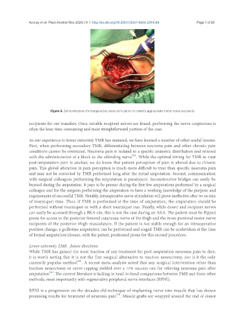
Figure 4. Demonstration of intraoperative nerve stimulation to identify appropriate motor nerve recipients
recipients for our transfers. Once suitable recipient nerves are found, performing the nerve coaptations is
often the least time consuming and most straightforward portion of the case.
As our experience in lower extremity TMR has matured, we have learned a number of other useful lessons.
First, when performing secondary TMR, differentiating between neuroma pain and other chronic pain
conditions cannot be overstated. Neuroma pain is isolated to a specific anatomic distribution and relieved
[48]
with the administration of a block to the offending nerve . While the optimal timing for TMR to treat
post-amputation pain is unclear, we do know that patient perception of pain is altered due to chronic
pain. This global alteration in pain perception is much more difficult to treat than specific neuroma pain
and may not be corrected by TMR performed long after the initial amputation. Second, communication
with surgical colleagues performing the amputation is paramount. Reconstructive bridges can easily be
burned during the amputation. It pays to be present during the first few amputations performed by a surgical
colleague and for the surgeon performing the amputation to have a working knowledge of the purpose and
requirements of successful TMR. Notably, intraoperative nerve stimulation will prove ineffective after 30-60 min
of tourniquet time. Thus, if TMR is performed at the time of amputation, the amputation should be
performed without tourniquet or with a short tourniquet run. Finally, while donor and recipient nerves
can easily be accessed through a BKA site, this is not the case during an AKA. The patient must be flipped
prone for access to the posterior femoral cutaneous nerve of the thigh and the more proximal motor nerve
recipients of the posterior thigh musculature. If the patient is not stable enough for an intraoperative
position change, a guillotine amputation can be performed and staged TMR can be undertaken at the time
of formal amputation/closure, with the patient positioned prone for this second procedure.
Lower extremity TMR - future directions
While TMR has gained the most traction of any treatment for post-amputation neuroma pain to date,
it is worth noting that it is not the first surgical alternative to traction neurectomy, nor is it the only
[49]
currently popular method . A recent meta-analysis noted that any surgical intervention other than
traction neurectomy or nerve capping yielded over a 75% success rate for relieving neuroma pain after
[50]
amputation . The current literature is lacking in head-to-head comparisons between TMR and these other
methods, most importantly with regenerative peripheral nerve interfaces (RPNI).
RPNI is a progression on the decades-old technique of implanting nerve into muscle that has shown
promising results for treatment of neuroma pain . Muscle grafts are wrapped around the end of donor
[51]