
Page 40 - Read Online
P. 40
Page 6 of 20 Azoury et al. Plast Aesthet Res 2020;7:4 I http://dx.doi.org/10.20517/2347-9264.2019.44
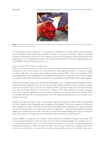
Figure 3. Dissection of a large neuroma in the common peroneal nerve of a transfemoral amputee with severe neuroma pain. Note the
haphazard organization of the neuroma
[45]
to communication between amputees . The majority of TMR that we currently perform at our institution
is in coordination with vascular and orthopedic surgeons at the time of amputation. This has re-framed
amputation from failure on the part of the surgeon and the end of the surgeon-patient relationship to the
beginnings of a new collaborative reconstructive relationship that results in less post-amputation pain and
the possibility of more advanced prosthetic use.
Lower extremity TMR - technical considerations
TMR in the lower extremity is commonly performed at the transfemoral level with above the knee
amputation (AKA) and transtibial level with below the knee amputation (BKA). In contrast to upper
extremity TMR, which can require more creativity, lower extremity TMR is often more formulaic. When
performing TMR for the management or prevention of neuroma pain, the specific motor nerve recipients
are much less important because they do not have to be superficial or separated from other nearby signals.
In the case of secondary neuroma treatment, the symptomatic nerve is isolated (usually through an incision
separate from the amputation site), the neuroma resected, and the fresh nerve end is coapted to a nearby
motor nerve recipient [Figure 3]. In the case of primary TMR, each pure sensory and mixed motor/sensory
nerve must be coapted directly to a motor nerve. During an AKA, this includes the posterior cutaneous
nerve of the thigh, the saphenous nerve, the common peroneal nerve, and the tibial nerve. During a BKA,
this includes the tibial nerve, the deep peroneal nerve, the superficial peroneal nerve, and the sural nerves
(medial and lateral).
Similar to the upper extremity, many of these initial surgical techniques and much of the continued data
for TMR originates from Dumanian and colleagues at Northwestern. They have populated the literature
with roadmaps for TMR at both the tranfemoral and transtibial levels, including the common locations
of frequently found motor nerves that can be used for coaptation [46,47] . We found these guides particularly
helpful in our initial forays into TMR. Since then, we have realized that finding motor nerves in practice is
much more reliant on intraoperative nerve stimulation than was first understood [Figure 4].
When available, we typically use a biphasic nerve stimulator (Checkpoint Surgical; Cleveland, OH)
because repeated stimulation with this system does not result in neuronal fatigue. The stimulator is set at
a moderate pulse duration and 2 mA for anterograde stimulation or 20 mA for retrograde stimulation.
Retrograde stimulation is often particularly useful. In fact, we have found that tracing proximal motor
nerve exit locations from a transected nerve is the most expeditious way to find suitable motor nerve