
Page 38 - Read Online
P. 38
Page 4 of 20 Azoury et al. Plast Aesthet Res 2020;7:4 I http://dx.doi.org/10.20517/2347-9264.2019.44
A B C
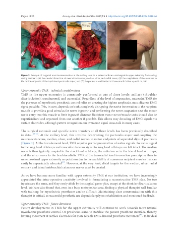
Figure 2. Example of targeted muscle reinnervation at the axillary level in a patient with an unsalvageable upper extremity from a drag
racing accident: (A) the careful dissection of musculocutaneous, median, ulnar, and radial nerve; (B) the coaptations of these nerves to
the motor endpoints of the ispsilateral pectoralis major; and (C) the patient is well healed at three-month follow up with no pain
Upper extremity TMR - technical considerations
TMR in the upper extremity is commonly performed at one of three levels: axillary (shoulder
disarticulation), transhumeral, and transradial. Regardless of the level of amputation, successful TMR for
the purposes of myoelectric prosthetic control relies on creating the highest amplitude, most discrete EMG
signal possible. This, in turn, depends on both completely disrupting the native innervation to the recipient
muscle to provide a good stimulus for nerve ingrowth and performing the nerve coaptation near the motor
nerve entry into this muscle to limit ingrowth distance. Recipient motor nerve/muscle units should also be
superficialized and separated from one another if possible. This allows easy decoding of EMG signals via
surface electrodes, although pattern recognition can overcome signal cross-talk in many cases.
The surgical rationale and specific nerve transfers at all three levels has been previously described
in detail [26-28] . At the axillary level, this involves denervating the pectoralis major and coapting the
musculocutaneous, median, ulnar, and radial nerves to motor endpoints of separated slips of pectoralis
[Figure 2]. At the transhumeral level, TMR requires partial preservation of native signals: the radial signal
to the long head of triceps and musculocutaneous signal to long head of biceps are left intact. The median
nerve is then typically coapted to the short head of biceps, the radial nerve to the lateral head of triceps,
and the ulnar nerve to the brachioradialis. TMR at the transradial level is even less prescriptive than in
more proximal upper extremity amputations due to the availability of numerous recipient muscles that can
[27]
easily be superficially relocated . However, at the very least, distal targets for the median, ulnar, radial
sensory, and lateral antebrachial cutaneous nerves must be created.
As we have become more familiar with upper extremity TMR at our institution, we have increasingly
appreciated the intra-operative creativity involved in formulating a reconstructive TMR plan. No two
injuries are the same, and they rarely abide by the surgical game-plan, except at the shoulder disarticulation
level. We have also found that, even in a busy metropolitan area, finding a physical therapist well familiar
with training for myoelectric prostheses can be difficult. Maintaining clear communication with this
therapist is critical, as successful prosthetic use depends largely on rehabilitation and monitored feedback.
Upper extremity TMR - future directions
Future developments in TMR for the upper extremity will continue to work towards more natural
myoelectric prosthetic control. OI prostheses stand to stabilize the patient-prosthetic interface, thereby
[29]
limiting movement at surface electrodes for more reliable EMG directed prosthetic movement . Individual