
Page 408 - Read Online
P. 408
Guagnano et al. Plast Aesthet Res 2020;7:37 I http://dx.doi.org/10.20517/2347-9264.2020.21 Page 7 of 10
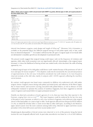
Table 4. Dental caries in primary (dmft) and permanent teeth (DMFT) by gender, ethnicity types of cleft, and surgical protocols in
the study sample (mean ± SD)
Variables dmft DMFT
Gender
Male (n = 51) 2.4 ± 2.9 0.9 ± 1.5
Female (n = 34) 1.8 ± 2.8 1.1 ± 2.1
Ethnicity
Caucasian (n = 68) 1.9 ± 2.9 1.8 ± 0.4
Asian (n = 7) 3.5 ± 2.6 0.4 ± 0.5
Hispanic (n = 5) 4.0 ± 2.9 1.0 ± 1.1
Indian (n = 1) 4.0 0.0
African (n = 4) 1 ± 1.1 1 ± 1.1
Cleft type
CLP (n = 64) 2.2 ± 1.0 1.1 ± 2.4
CP (n =10) 2.5 ± 1.0 0.3 ± 0.9
CLA (n =11) 1.5 ± 0.9 0.7 ± 1.0
Surgical protocol
EPP (n = 21) 3.9 ± 3.0* 0.8 ± 1.2
DPP (n = 61) 1.5 ± 2.6 1.1 ± 1.9
Values with superscript asterisks show statistically significant difference between groups: *P < 0.05. CLP: lip palate cleft; CP: palate cleft;
CLA: lip alveolar cleft; EPP: early palate periosteal plastic; DPR: delayed palate plastic
[6]
interval time between surgeries, study design and length of follow-up . Moreover, little information is
available on the potential effects that different surgical timings of hard palate repair, early or late, could
have on dental development [14,15] . It is indeed well known that all types of surgical repair of oro-facial clefts
are detrimental to maxillary growth and development of permanent teeth.
The present results suggest that surgical timing could impact only on the frequency of rotations and
agenesis, while other tooth anomalies were not significantly affected. Interestingly, a late surgical repair
increased 3.5 times the likelihood of having at least one rotated tooth, while decreased the odds of having
agenesis by 70%.
A delayed surgical repair of the cleft palate could favour tooth rotation because of the persistence of lack of
[18]
bone and space for incisor eruption . This situation could even be worsened by the concomitant presence
of supernumeraries in the cleft area. It should be considered that tooth rotation is the most frequently
observed anomaly at the cleft side, mainly in subjects with L-UCLP, especially affecting the maxillary
central incisors.
By contrast, tooth agenesis was found more commonly in children who underwent early cleft palate
surgical closure. Surgical trauma during early palatal periosteal plastic and reduction of the blood supply
due to tissue tension and excessive scarring associated with palatal defects, as well as absence of early
orthopaedic treatment to optimise the position of maxillary fragments, have been suggested as external
[14]
causes of agenesis and enamel defects in upper permanent incisors .
Notably, we observed a prevalence of tooth agenesis of 42.2%, that was lower than that reported in the
literature, ranging from 45% to 67.6% [19-23] . This lower frequency could be explained by the fact that most
of the patients were Caucasian, an ethnicity with a low incidence of agenesis, and underwent a late surgical
closure of the hard palate, at 4 years of age or older. Tooth agenesis affected more frequently BCLP subjects
(71.4%), in which the alveolar defect is more severe than in other cleft types. According to the literature,
upper lateral incisors were the more common missing teeth (40%), followed by upper and lower second
[6]
premolars (18%), lower left incisors and lower second molars (2%) .
Morphological abnormalities of dental crowns were observed in 20.5% of the patients, being the lateral
incisors the most frequently affected teeth. In particular, microdontia was found in 14.5% of cleft children,