
Page 352 - Read Online
P. 352
Costa et al. Plast Aesthet Res 2020;7:32 I http://dx.doi.org/10.20517/2347-9264.2020.43 Page 9 of 12
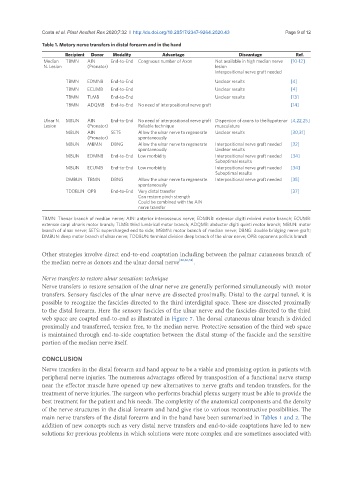
Table 1. Motory nerve transfers in distal forearm and in the hand
Recipient Donor Modality Advantage Disvantage Ref.
Median TBMN AIN End-to-End Congruous number of Axon Not available in high median nerve [10-12]
N. Lesion (Pronator) lesion
Interpositional nerve graft needed
TBMN EDMNB End-to-End Unclear results [4]
TBMN ECUMB End-to-End Unclear results [4]
TBMN TLMB End-to-End Unclear results [13]
TBMN ADQMB End-to-End No need of interpositional nerve graft [14]
Ulnar N. MBUN AIN End-to-End No need of interpositional nerve graft Dispersion of axons to the hypotenar [4,22,25]
Lesion (Pronator) Reliable technique musculature
MBUN AIN SETS Allow the ulnar nerve to regenerate Unclear results [30,31]
(Pronator) spontaneously
MBUN MBMN DBNG Allow the ulnar nerve to regenerate Interpositional nerve graft needed [32]
spontaneously Unclear results
MBUN EDMNB End-to-End Low morbidity Interpositional nerve graft needed [34]
Suboptimal results
MBUN ECUMB End-to-End Low morbidity Interpositional nerve graft needed [34]
Suboptimal results
DMBUN TBMN DBNG Allow the ulnar nerve to regenerate Interpositional nerve graft needed [35]
spontaneously
TDDBUN OPB End-to-End Very distal transfer [37]
Can restore pinch strength
Could be combined with the AIN
nerve transfer
TBMN: Thenar branch of median nerve; AIN: anterior interosseous nerve; EDMNB: extensor digiti minimi motor branch; ECUMB:
extensor carpi ulnaris motor branch; TLMB: third lumbrical motor branch; ADQMB: abductor digiti quinti motor branch; MBUN: motor
branch of ulnar nerve; SETS: supercharged end to side; MBMN: motor branch of median nerve; DBNG: double bridging nerve graft;
DMBUN: deep motor branch of ulnar nerve; TDDBUN: terminal division deep branch of the ulnar nerve; OPB: opponens pollicis branch
Other strategies involve direct end-to-end coaptation including between the palmar cutaneous branch of
the median nerve as donors and the ulnar dorsal nerve [22,56,58] .
Nerve transfers to restore ulnar sensation: technique
Nerve transfers to restore sensation of the ulnar nerve are generally performed simultaneously with motor
transfers. Sensory fascicles of the ulnar nerve are dissected proximally. Distal to the carpal tunnel, it is
possible to recognize the fascicles directed to the third interdigital space. These are dissected proximally
to the distal forearm. Here the sensory fascicles of the ulnar nerve and the fascicles directed to the third
web space are coapted end-to-end as illustrated in Figure 7. The dorsal cutaneous ulnar branch is divided
proximally and transferred, tension free, to the median nerve. Protective sensation of the third web space
is maintained through end-to-side coaptation between the distal stump of the fascicle and the sensitive
portion of the median nerve itself.
CONCLUSION
Nerve transfers in the distal forearm and hand appear to be a viable and promising option in patients with
peripheral nerve injuries. The numerous advantages offered by transposition of a functional nerve stump
near the effector muscle have opened up new alternatives to nerve grafts and tendon transfers, for the
treatment of nerve injuries. The surgeon who performs brachial plexus surgery must be able to provide the
best treatment for the patient and his needs. The complexity of the anatomical components and the density
of the nerve structures in the distal forearm and hand give rise to various reconstructive possibilities. The
main nerve transfers of the distal forearm and in the hand have been summarized in Tables 1 and 2. The
addition of new concepts such as very distal nerve transfers and end-to-side coaptations have led to new
solutions for previous problems in which solutions were more complex and are sometimes associated with