
Page 349 - Read Online
P. 349
Page 6 of 12 Costa et al. Plast Aesthet Res 2020;7:32 I http://dx.doi.org/10.20517/2347-9264.2020.43
Figure 4. The fourth digital nerve is transferred end-to-end to the first digital nerve. The remaining median-dependent distal stumps are
coapted end-to-side. Yellow: functional nerves; lighter yellow: sensitive areas; pink: nonfunctional nerves
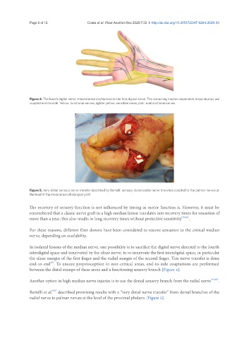
Figure 5. Very distal sensory nerve transfer described by Bertelli: sensory dorsal radial nerve branches coapted to the palmar nerves at
the level of the metacarpal-phalangeal joint
The recovery of sensory function is not influenced by timing as motor function is. However, it must be
remembered that a classic nerve graft in a high median lesion translates into recovery times for sensation of
more than a year; this also results in long recovery times without protective sensitivity [39,40] .
For these reasons, different fiber donors have been considered to restore sensation to the critical median
nerve, depending on availability.
In isolated lesions of the median nerve, one possibility is to sacrifice the digital nerve directed to the fourth
interdigital space and innervated by the ulnar nerve, to re-innervate the first interdigital space, in particular
the ulnar margin of the first finger and the radial margin of the second finger. This nerve transfer is done
[4]
end-to-end . To ensure proprioception in non-critical areas, end-to-side coaptations are performed
between the distal stumps of these areas and a functioning sensory branch [Figure 4].
Another option in high median nerve injuries is to use the dorsal sensory branch from the radial nerve [41,42] .
[43]
Bertelli et al. described promising results with a “very distal nerve transfer” from dorsal branches of the
radial nerve to palmar nerves at the level of the proximal phalanx [Figure 5].