
Page 31 - Read Online
P. 31
Page 4 of 7 Kishi. Plast Aesthet Res 2020;7:3 I http://dx.doi.org/10.20517/2347-9264.2019.50
A B
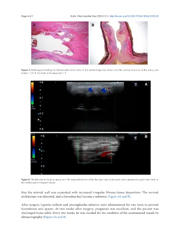
Figure 4. Pathological findings by Hematoxylin-Eosin stain A: the hemorrhage was shown and the normal structure of the artery was
broken × 20; B: the neck of the aneurysm × 4
A
B
Figure 5. The left side is the ulnar artery and The longitudinal view of the the right side is the radial artery anastomotic graft vein (red). at
the middle part of the palm (blue)
that the arterial wall was expanded with increased irregular fibrous tissue deposition. The normal
architecture was distorted, and a thrombus had become a substrate [Figure 4A and B].
After surgery, heparin sodium and prostaglandin infusion were administered for one week to prevent
thrombosis and spasm. At two weeks after surgery, prognosis was excellent, and the patient was
discharged home safely. Every two weeks, he was checked for the condition of the anastomosed vessels by
ultrasonography [Figure 5A and B].