
Page 29 - Read Online
P. 29
Page 2 of 7 Kishi. Plast Aesthet Res 2020;7:3 I http://dx.doi.org/10.20517/2347-9264.2019.50
B
A
C D
E F G
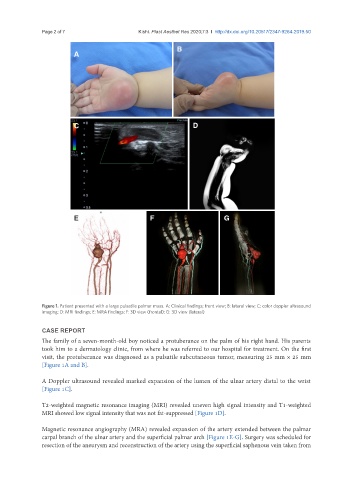
Figure 1. Patient presented with a large pulsatile palmar mass. A: Clinical findings; front view; B: lateral view; C: color doppler ultrasound
imaging; D: MRI findings; E: MRA findings; F: 3D view (frontal); G: 3D view (lateral)
CASE REPORT
The family of a seven-month-old boy noticed a protuberance on the palm of his right hand. His parents
took him to a dermatology clinic, from where he was referred to our hospital for treatment. On the first
visit, the protuberance was diagnosed as a pulsatile subcutaneous tumor, measuring 25 mm × 25 mm
[Figure 1A and B].
A Doppler ultrasound revealed marked expansion of the lumen of the ulnar artery distal to the wrist
[Figure 1C].
T2-weighted magnetic resonance imaging (MRI) revealed uneven high signal intensity and T1-weighted
MRI showed low signal intensity that was not fat-suppressed [Figure 1D].
Magnetic resonance angiography (MRA) revealed expansion of the artery extended between the palmar
carpal branch of the ulnar artery and the superficial palmar arch [Figure 1E-G]. Surgery was scheduled for
resection of the aneurysm and reconstruction of the artery using the superficial saphenous vein taken from