
Page 30 - Read Online
P. 30
Kishi. Plast Aesthet Res 2020;7:3 I http://dx.doi.org/10.20517/2347-9264.2019.50 Page 3 of 7
A B
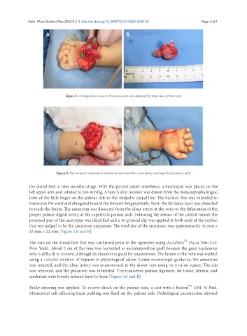
Figure 2. Intraoperative view A: the aneurysm was elevated; B: total view of the mass
A B
Figure 3. The recipient vein was anastomosis between the ulnar artery and superficial palmar arch
the dorsal foot at nine months of age. With the patient under anesthesia, a tourniquet was placed on the
left upper arm and inflated to 200 mmHg. A lazy-S skin incision was drawn from the metacarpophalangeal
joint of the little finger on the palmar side to the midpalm carpal line. The incision line was extended to
transverse the wrist and elongated toward the forearm longitudinally. Next, the fat tissue layer was dissected
to reach the lesion. The aneurysm was dissected from the ulnar artery at the wrist to the bifurcation of the
proper palmar digital artery at the superficial palmar arch. Following the release of the cubital tunnel, the
proximal part of the aneurysm was identified and a 30-g vessel clip was applied to both ends of the section
that was judged to be the aneurysm expansion. The total size of the aneurysm was approximately 32 mm ×
25 mm × 22 mm [Figure 2A and B].
TX
The vain on the dorsal foot that was confirmed prior to the operation using AccuVein (Accu Vein LLC
New York). About 2 cm of the vein was harvested as an interposition graft because the great saphenous
vein is difficult to remove, although its diameter is good for anastomosis. The lumen of the vein was washed
using a 1:20,000 solution of heparin in physiological saline. Under microscopic guidance, the aneurysm
was resected, and the ulnar artery was anastomosed to the donor vein using 10-0 nylon suture. The clip
was removed, and the pulsation was identified. The transverse palmar ligament, fat tissue, dermis, and
epidermis were loosely sutured layer by layer [Figure 3A and B].
TM
Bulky dressing was applied. To relieve shock on the palmar side, a cast with a Reston (3M. St Paul.
Minnesota) self-adhering foam padding was fixed on the palmar side. Pathological examination showed