Page 266 - Read Online
P. 266
Ramirez. Plast Aesthet Res 2020;7:25  I  http://dx.doi.org/10.20517/2347-9264.2019.78                                               Page 7 of 21


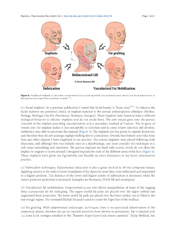
               Figure 6. Additional methods of volumetric enhancement that can be applied to the pureendoscopic central oval facial rejuvenation or
               the biplanar technique (First published in study [22] )

                                                                                          [36]
               (1) Facial implants. In a previous publication I stated that facial beauty is “bone deep” . To enhance the
               facial skeleton my preferred choice of implant material is the porous polypropylene [Medpor (Stryker.
               Portage, Michigan) Su-Por (Poriferous. Newman, Georgia)]. These implants were found to have a different
               biological behavior to silicone implants and do not erode bone. The soft tissues grew into the porous
               channels of the implant providing vascularization and a secondary method of fixation. The in-grow of
               vessels into the implant makes it less susceptible to infection and in cases where infection did develop,
               antibiotics were able to penetrate the implant [Figure 7]. The implants are less prone to capsule formation,
               and therefore they do not undergo implant shifting due to contracture. Overall, they behave more like bone
               than any other implant I have employed in my practice. The porous implants were placed following wide
               dissection, and although this was initially seen as a disadvantage, one must consider this technique as a
               soft tissue remodeling and reposition. The porous implants are fixed with screws, which do not allow the
               implant to migrate or move around. I designed implants for each of the different areas of the face [Figure 8].
               These implants have given me figuratively and literally an extra dimension to my facial rejuvenation
               practice.


               (2) Imbrication techniques. Subperiosteal dissection is also a great method to lift the composite tissues.
               Applying sutures to the mid or lower boundaries of the dissected areas they were imbricated and suspended
               to a higher position. The distance of the lower and higher points of imbrication is decreased while the
               antero-posterior projection is increased. Examples are browpexy, SOOF lift and mentopexy.

               (3) Vascularized fat mobilization. Subperiosteal access also allows manipulation of some of the sagging
               deep scyssarcosis fat for reshaping. The upper eyelid fat pads are placed over the upper orbital rim
               augmented brow projection. The lower eyelid fat pads are placed over the lower orbital rim to filled in the
               tear trough region. The herniated Bichat’s fat pad is used to create the Ogee line of the midface.


               (4) Fat grafting. With subperiosteal endoscopic techniques there is no associated delamination of the
               anatomical planes, therefore fat can be injected anywhere from dermis to periosteum. Fat is injected with
               1cc Luer-Lock syringes attached to the “Ramirez Super-Luer-Lock micro-cannulas” (Tulip Medical, San

