
Page 263 - Read Online
P. 263
Page 4 of 21 Ramirez. Plast Aesthet Res 2020;7:25 I http://dx.doi.org/10.20517/2347-9264.2019.78
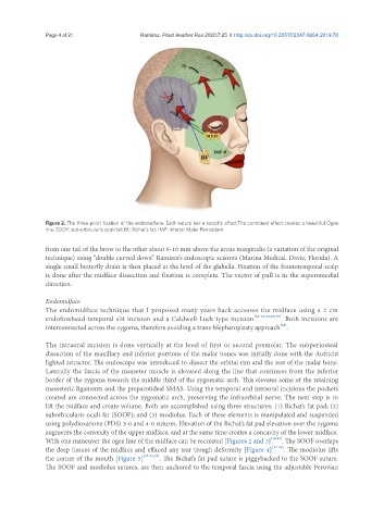
Figure 2. The three-point fixation of the endomidface. Each suture has a specific effect.The combined effect creates a beautiful Ogee
line. SOOF: sub-orbicularis oculi fat; BF: Bichat’s fat; IMP: inferior Malar Periostium
from one tail of the brow to the other about 5-10 mm above the arcus marginalis (a variation of the original
technique) using “double curved down” Ramirez’s endoscopic scissors (Marina Medical. Davie, Florida). A
single small butterfly drain is then placed at the level of the glabella. Fixation of the frontotemporal scalp
is done after the midface dissection and fixation is complete. The vector of pull is in the superomedial
direction.
Endomidface
The endomidface technique that I proposed many years back accesses the midface using a 2 cm
endoforehead-temporal slit incision and a Caldwell-Luck type incision [22-24,28,29,33] . Both incisions are
[34]
interconnected across the zygoma, therefore avoiding a trans-blepharoplasty approach .
The intraoral incision is done vertically at the level of first or second premolar. The subperiosteal
dissection of the maxillary and inferior portions of the malar bones was initially done with the Aufricht
lighted retractor. The endoscope was introduced to dissect the orbital rim and the rest of the malar bone.
Laterally the fascia of the masseter muscle is elevated along the line that continues from the inferior
border of the zygoma towards the middle third of the zygomatic arch. This elevates some of the retaining
masseteric ligaments and the preparotideal SMAS. Using the temporal and intraoral incisions the pockets
created are connected across the zygomatic arch, preserving the infraorbital nerve. The next step is to
lift the midface and create volume. Both are accomplished using three structures: (1) Bichat’s fat pad; (2)
suborbicularis oculi fat (SOOF); and (3) modiolus. Each of these elements is manipulated and suspended
using polydioxanone (PDS) 3-0 and 4-0 sutures. Elevation of the Bichat’s fat pad elevation over the zygoma
augments the convexity of the upper midface, and at the same time creates a concavity of the lower midface.
With one maneuver the ogee line of the midface can be recreated [Figures 2 and 3] [29,33] . The SOOF overlaps
the deep tissues of the midface and effaced any tear trough deformity [Figure 4] [17-25] . The modiolus lifts
the corner of the mouth [Figure 5] [22-24,33] . The Bichat’s fat pad suture is piggybacked to the SOOF suture.
The SOOF and modiolus sutures, are then anchored to the temporal fascia using the adjustable Peruvian