
Page 262 - Read Online
P. 262
Ramirez. Plast Aesthet Res 2020;7:25 I http://dx.doi.org/10.20517/2347-9264.2019.78 Page 3 of 21
C 1996 Oscar Ramirez MD
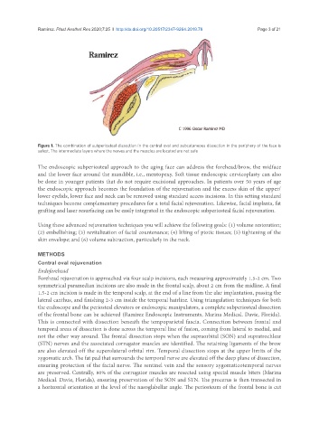
Figure 1. The combination of subperiosteal dissection in the central oval and subcutaneous dissection in the periphery of the face is
safest. The intermediate layers where the nerves and the muscles are located are not safe
The endoscopic subperiosteal approach to the aging face can address the forehead/brow, the midface
and the lower face around the mandible, i.e., mentopexy. Soft tissue endoscopic cervicoplasty can also
be done in younger patients that do not require excisional approaches. In patients over 50 years of age
the endoscopic approach becomes the foundation of the rejuvenation and the excess skin of the upper/
lower eyelids, lower face and neck can be removed using standard access incisions. In this setting standard
techniques become complementary procedures for a total facial rejuvenation. Likewise, facial implants, fat
grafting and laser resurfacing can be easily integrated in the endoscopic subperiosteal facial rejuvenation.
Using these advanced rejuvenation techniques you will achieve the following goals: (1) volume restoration;
(2) embellishing; (3) revitalization of facial countenance; (4) lifting of ptotic tissues; (5) tightening of the
skin envelope; and (6) volume subtraction, particularly in the neck.
METHODS
Central oval rejuvenation
Endoforehead
Forehead rejuvenation is approached via four scalp incisions, each measuring approximately 1.5-2 cm. Two
symmetrical paramedian incisions are also made in the frontal scalp, about 2 cm from the midline. A final
1.5-2 cm incision is made in the temporal scalp, at the end of a line from the alar implantation, passing the
lateral canthus, and finishing 2-3 cm inside the temporal hairline. Using triangulation techniques for both
the endoscope and the periosteal elevators or endoscopic manipulators, a complete subperiosteal dissection
of the frontal bone can be achieved (Ramirez Endoscopic Instruments, Marina Medical. Davie, Florida).
This is connected with dissection beneath the tempoparietal fascia. Connection between frontal and
temporal areas of dissection is done across the temporal line of fusion, coming from lateral to medial, and
not the other way around. The frontal dissection stops when the supraorbital (SON) and supratrochlear
(STN) nerves and the associated corrugator muscles are identified. The retaining ligaments of the brow
are also elevated off the superolateral orbital rim. Temporal dissection stops at the upper limits of the
zygomatic arch. The fat pad that surrounds the temporal nerve are elevated off the deep plane of dissection,
ensuring protection of the facial nerve. The sentinel vein and the sensory zygomaticotemporal nerves
are preserved. Centrally, 80% of the corrugator muscles are resected using special muscle biters (Marina
Medical. Davie, Florida), ensuring preservation of the SON and STN. The procerus is then transected in
a horizontal orientation at the level of the nasoglabellar angle. The periosteum of the frontal bone is cut