
Page 123 - Read Online
P. 123
Campbell et al. Plast Aesthet Res 2020;7:12 I http://dx.doi.org/10.20517/2347-9264.2019.59 Page 3 of 6
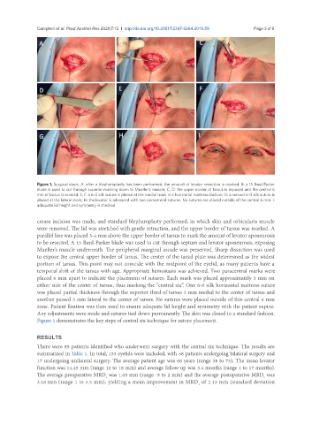
Figure 1. Surgical steps. A: after a blepharoplasty has been performed, the amount of levator resection is marked; B: a 15 Bard-Parker
blade is used to cut through superior marking down to Mueller’s muscle; C, D: the upper border of tarsus is exposed and the central 6
mm of tarsus is marked; E, F: a 6-0 silk suture is placed at the medial mark in a horizontal mattress fashion; G: a second 6-0 silk suture is
placed at the lateral mark; H: the levator is advanced with two paracentral sutures. No sutures are placed outside of the central 6 mm; I:
adequate lid height and symmetry is checked
crease incision was made, and standard blepharoplasty performed, in which skin and orbicularis muscle
were removed. The lid was stretched with gentle retraction, and the upper border of tarsus was marked. A
parallel line was placed 3-4 mm above the upper border of tarsus to mark the amount of levator aponeurosis
to be resected. A 15 Bard-Parker blade was used to cut through septum and levator aponeurosis, exposing
Mueller’s muscle underneath. The peripheral marginal arcade was preserved. Sharp dissection was used
to expose the central upper border of tarsus. The center of the tarsal plate was determined as the widest
portion of tarsus. This point may not coincide with the midpoint of the eyelid, as many patients have a
temporal shift of the tarsus with age. Appropriate hemostasis was achieved. Two paracentral marks were
placed 6 mm apart to indicate the placement of sutures. Each mark was placed approximately 3 mm on
either side of the center of tarsus, thus marking the “central six”. One 6-0 silk horizontal mattress suture
was placed partial thickness through the superior third of tarsus 3 mm medial to the center of tarsus and
another passed 3 mm lateral to the center of tarsus. No sutures were placed outside of this central 6 mm
zone. Patient fixation was then used to ensure adequate lid height and symmetry with the patient supine.
Any adjustments were made and sutures tied down permanently. The skin was closed in a standard fashion.
Figure 1 demonstrates the key steps of central six technique for suture placement.
RESULTS
There were 85 patients identified who underwent surgery with the central six technique. The results are
summarized in Table 1. In total, 153 eyelids were included, with 68 patients undergoing bilateral surgery and
17 undergoing unilateral surgery. The average patient age was 68 years (range 38 to 73). The mean levator
function was 14.45 mm (range 12 to 18 mm) and average follow-up was 3.4 months (range 1 to 17 months).
The average preoperative MRD was 1.05 mm (range -5 to 2 mm) and the average postoperative MRD was
1
1
3.18 mm (range 1 to 5.5 mm), yielding a mean improvement in MRD of 2.13 mm (standard deviation
1