
Page 244 - Read Online
P. 244
Nicholson et al. Plast Aesthet Res 2018;5:34 I http://dx.doi.org/10.20517/2347-9264.2018.30 Page 7 of 11
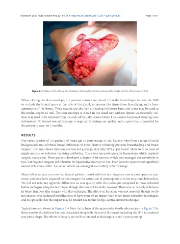
Figure 6. Plication of the inferior dermal strip to shorten the distance between the nipple and the inframammary fold
When closing the skin envelope, 2-0 prolene sutures are placed from the fascial layer to near the IMF
to occlude the lateral space at the side of the gland, to prevent the tissue from lateralising and a boxy
appearance of the breast. These sutures are also key to shaping the breast base, and some may be used at
the medial aspect as well. The skin envelope is closed in the usual way without drains. Occasionally, vol-
ume may need to be removed from the ends of the IMF wound before final closure to prevent standing cone
deformities. No formal wound drainage is required. Dressings are applied and a sports bra is provided for
the patient to wear for 2 months.
RESULTS
Our series consists of 116 patients, of mean age 40 years (range 19-65). Patients were from a range of racial
backgrounds and exhibited broad differences in breast history including previous breastfeeding and breast
surgery. The mean tissue mass excised was 865 g (range: skin only-2175 g) per breast. There were no cases of
nipple necrosis or infection requiring antibiotics. There was one post-operative haematoma which required
surgical evacuation. Three patients developed a degree of fat necrosis which was managed conservatively in
two, but required surgical debridement for liquefactive necrosis in one. Four patients experienced superficial
wound dehiscence at the T-junction which was managed successfully with dressings.
Mean follow up was 20.6 months. Several patients treated with this technique are now at post-operative year
seven, and none have required revision surgery for correction of pseudoptosis or other recurrent deformities.
We did not note any apparent differences in scar quality with this technique compared to those obtained
before we began using the technique, though this was not formally assessed. There was no notable difference
in breast firmness after surgery with this technique. The effects on lactation were not assessed, though we do
not expect these technical modifications to have more of an impact than other breast reduction techniques,
and it is possible that the impact may be smaller due to this being a central mound technique.
Typical cases are shown in Figures 7-9. Note the fullness at the upper poles shortly after surgery in Figure 7. By
three months this fullness has now descended along with the rest of the breast, including the IMF to a natural
non-ptotic shape. The effects of surgery are well-maintained at discharge at 2 and 3 years post-op.