
Page 241 - Read Online
P. 241
Page 4 of 11 Nicholson et al. Plast Aesthet Res 2018;5:34 I http://dx.doi.org/10.20517/2347-9264.2018.30
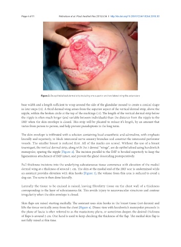
Figure 2. De-epithelialised dermal strip including one superior and two lateral wing-like extensions
base width and a length sufficient to wrap around the side of the glandular mound to create a conical shape
in later steps (11). A third dermal wing arises from the superior aspect of the vertical dermal strip, above the
nipple, within the broken circle at the top of the markings (12). The length of the vertical dermal strip below
the nipple is often much longer (and variable between individuals) than the distance from the nipple to the
IMF when the skin envelope is closed. This strip will be plicated to reduce it’s length, by an amount that
varies from person to person, and help prevent pseudoptosis in the long term.
The skin envelope is infiltrated with a solution containing local anaesthetic and adrenaline, with emphasis
laterally and superiorly, to block intercostal nerve sensory branches and constrict the intercostal perforator
vessels. The smaller breast is reduced first. All of the marks are scored. Without the use of a breast
tourniquet, the vertical dermal strip, along with the 3 dermal “wings”, are de-epithelialised using handswitch
monopolar, sparing the nipple [Figure 2]. The incision parallel to the IMF is beveled superiorly to keep the
ligamentous attachment of IMF intact, and prevent the gland descending postoperatively.
Full thickness incisions into the underlying subcutaneous tissue commence with elevation of the medial
dermal wing at a thickness of around 1 cm. The skin at the medial end of the IMF scar is undermined while
an assistant provides elevation with skin hooks [Figure 3]; the volume from this area is reduced to avoid a
dog-ear. The same is then done laterally.
Laterally the tissue to be excised is raised, leaving fibrofatty tissue on the chest wall of a thickness
corresponding to the layer of subcutaneous fat. This avoids injury to neurovascular structures and contour
irregularity when the skin envelope is closed.
Skin flaps are raised starting medially. The assistant uses skin hooks in the breast tissue (not dermis) and
lifts the tissue vertically away from the chest [Figure 4]. Dissec-tion with handswitch monopolar proceeds in
the plane of fascia is often referred to as the mastectomy plane, or sometimes deeper; the desired thickness
of flaps is around 2 cm. One hand is used to keep checking the thickness of the flap. The medial skin flap is
not fully raised at this time.