
Page 240 - Read Online
P. 240
Nicholson et al. Plast Aesthet Res 2018;5:34 I http://dx.doi.org/10.20517/2347-9264.2018.30 Page 3 of 11
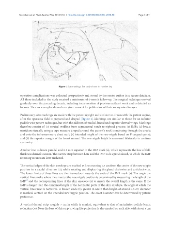
Figure 1. Skin markings. See body of text for number key
operative complications was collected prospectively and stored by the senior author in a secure database.
All those included in the study received a minimum of 6-month follow-up. The surgical technique evolved
gradually over the preceding decade, including incorporation of previous authors’ work and is detailed as
follows. The case examples shown have given consent for publication of their anonymised images.
Preliminary skin markings are made with the patient upright and are later re-drawn with the patient supine,
after the operative field is prepared and draped [Figure 1]. Markings are similar to those for an inferior
pedicle wise pattern technique, but with the addition of medial, lateral and superior dermal wings. Markings
therefore consist of: (1) vertical midline from suprasternal notch to xiphoid process; (2) IMFs; (3) breast
meridians (usually using a tape measure draped around the patient’s neck) continuing through the areola
and onto the inframammary chest wall; (4) intended height of the new nipple based on Pitanguy’s point;
and (5) the superior margin of the breast mound. The new nipple height is measured bilaterally to confirm
symmetry.
Another line is drawn parallel and 2-3 mm superior to the IMF mark (6), which represents the line of full-
thickness dermal incision. The narrow strip between here and the IMF is de-epithelialised, to which the IMF
retaining sutures are later anchored.
The vertical edges of the skin envelope are marked as lines running 7.5 cm from the centre of the new nipple
position in a caudal direction (7), while rotating and displac-ing the gland clockwise and anticlockwise.
The lower limits of these lines are then turned 90º towards the ends of the IMF mark (8). The angle the
vertical lines make where they meet at the new nipple position is determined by measuring the length of the
[2]
IMF and the corresponding lines of the skin envelope (8) to ensure the overall length is the same. If the
IMF is longer than the combined length of the horizontal parts of the skin envelope, the angle at which the
vertical lines meet is narrowed. A broken circle (9), greater in width than height, of around 4.5 cm diameter
is marked, centred on the intended new nipple position. The exact diameter can be determined by patient
preference.
A vertical dermal strip roughly 7 cm in width is marked, equivalent to that of an inferior pedicle breast
reduction (10). Near the base of this strip, a wing-like projection is also marked on each side, with about 5 cm