
Page 198 - Read Online
P. 198
Weber et al. Pressure ulcer coverage with contralateral gracilis
Unfortunately, despite adequate flap coverage,
nearly one-third of patients will develop a recurrent
pressure ulcer [4,5] . A history of previous pressure ulcer
increases the risk of developing a second pressure
ulcer, particularly in the ischial region [1,6] . Fortunately,
many of the flaps commonly used for pressure ulcer
treatment can be readvanced or reused under certain
conditions. However, difficulty arises when the patient
requires repeated coverage of large ulcers in the
same location and has exhausted the available local
muscle options. Here, we present a case of recurrent
ischioperineal ulcer in which the contralateral gracilis
muscle was used for wound coverage.
CASE REPORT
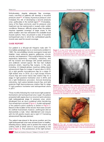
Our patient is a 49-year-old Hispanic male with T3
incomplete paraplegia due to a motorcycle accident in Figure 1: A view of the right ischioperineal ulcer with the patient
1984. His comorbidities include neurogenic bowel and in the prone position. Note the multiple previous incisions.
bladder, lower extremity spasms, gallstones, chronic The planned left gracilis muscle flap and right V-Y hamstring
myocutaneous advancement flap were marked preoperatively
anemia, and depression. Past surgical history includes
exploratory laparotomy, orchiopexy, colostomy, and
left hip incision and drainage with partial osteotomy
and antibiotic cement spacer. He has had multiple
pressure ulcers, requiring flap surgery, in the past,
including: (1) bilateral gluteus maximus sliding island
advancement flaps for a stage IV sacral ulcer in 2010;
(2) a right gracilis myocutaneous flap for a stage IV
right ischial ulcer in 2014; (3) a right biceps femoris
muscle flap and tensor fascia lata rotation flap for a
stage IV right posterior trochanter ulcer in 2016; and
(4) a right Girdlestone procedure, femoral shortening
osteotomy, right vastus lateralis muscle flap, and right
posterior thigh fasciocutaneous rotation flap for stage
IV right posterior trochanter and ischioperineal ulcers Figure 2: Antro posterior (AP) radiograph of the patient’s pelvis
demonstrates rotation, bony resorption, and heterotopic ossification
in 2017. of the pelvis. History of a right Girdlestone procedure and
shortening of the right femur is evident. The left femoral head has
Three months following the most recent right posterior been resected and antibiotic cement spacer was placed in the left
acetabulum for treatment of a prior infection
trochanteric and ischioperineal ulcer repair, the patient
presented with sepsis due to a recurrent and extensive
right ischioperineal stage IV pressure ulcer, which
developed from an injury sustained while transferring
from wheelchair to shower [Figure 1]. A plain radiograph
of the patient’s pelvic anatomy is shown in Figure 2.
Based on the location of the wound, there was concern
for urethral involvement, however, urethrogram did not
demonstrate a leak. Ulcer debridement and closure
with the left gracilis muscle and a right V-Y hamstring
advancement flap was planned.
The patient was placed in the prone position and the
right ischioperineal ulcer was excised down to healthy,
bleeding tissue, taking care to protect the rectum and Figure 3: The right ischioperineal ulcer was debrided down to
urethra, both of which were in close proximity to the healthy bleeding tissue and the prominence of the ischial bone was
reduced. Note the proximity of the ulcer to the anus and extension
ulcer [Figure 3]. The prominence of the right ischium into the perineal region
Plastic and Aesthetic Research ¦ Volume 4 ¦ October 31, 2017 191