
Page 199 - Read Online
P. 199
Weber et al. Pressure ulcer coverage with contralateral gracilis
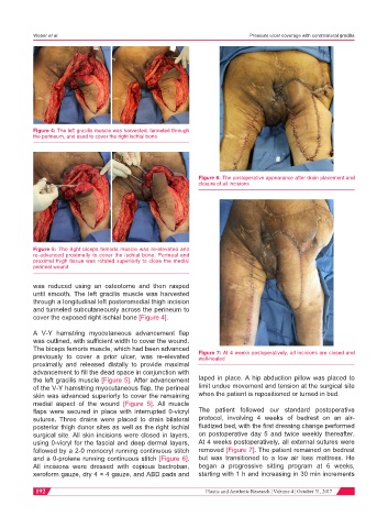
Figure 4: The left gracilis muscle was harvested, tunneled through
the perineum, and used to cover the right ischial bone
Figure 6: The postoperative appearance after drain placement and
closure of all incisions
Figure 5: The right biceps femoris muscle was re-elevated and
re-advanced proximally to cover the ischial bone. Perineal and
proximal thigh tissue was rotated superiorly to close the medial
perineal wound
was reduced using an osteotome and then rasped
until smooth. The left gracilis muscle was harvested
through a longitudinal left posteromedial thigh incision
and tunneled subcutaneously across the perineum to
cover the exposed right ischial bone [Figure 4].
A V-Y hamstring myocutaneous advancement flap
was outlined, with sufficient width to cover the wound.
The biceps femoris muscle, which had been advanced
previously to cover a prior ulcer, was re-elevated Figure 7: At 4 weeks postoperatively, all incisions are closed and
well-healed
proximally and released distally to provide maximal
advancement to fill the dead space in conjunction with
the left gracilis muscle [Figure 5]. After advancement taped in place. A hip abduction pillow was placed to
of the V-Y hamstring myocutaneous flap, the perineal limit undue movement and tension at the surgical site
skin was advanced superiorly to cover the remaining when the patient is repositioned or turned in bed.
medial aspect of the wound [Figure 5]. All muscle
flaps were secured in place with interrupted 0-vicryl The patient followed our standard postoperative
sutures. Three drains were placed to drain bilateral protocol, involving 4 weeks of bedrest on an air-
posterior thigh donor sites as well as the right ischial fluidized bed, with the first dressing change performed
surgical site. All skin incisions were closed in layers, on postoperative day 5 and twice weekly thereafter.
using 0-vicryl for the fascial and deep dermal layers, At 4 weeks postoperatively, all external sutures were
followed by a 2-0 monocryl running continuous stitch removed [Figure 7]. The patient remained on bedrest
and a 0-prolene running continuous stitch [Figure 6]. but was transitioned to a low air loss mattress. He
All incisions were dressed with copious bactroban, began a progressive sitting program at 6 weeks,
xeroform gauze, dry 4 × 4 gauze, and ABD pads and starting with 1 h and increasing in 30 min increments
192 Plastic and Aesthetic Research ¦ Volume 4 ¦ October 31, 2017