
Page 290 - Read Online
P. 290
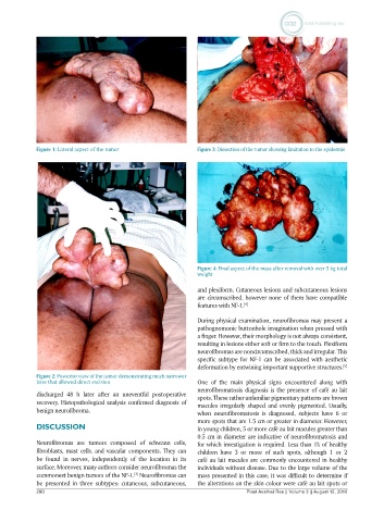
Figure 1: Lateral aspect of the tumor             Figure 3: Dissection of the tumor showing limitation to the epidermis


                                                              Figure 4: Final aspect of the mass after removal with over 5 kg total
                                                              weight

                                                              and plexiform. Cutaneous lesions and subcutaneous lesions
                                                              are circumscribed, however none of them have compatible
                                                              features with NF-1. [4]

                                                              During physical examination, neurofibromas may present a
                                                              pathognomonic buttonhole invagination when pressed with
                                                              a finger. However, their morphology is not always consistent,
                                                              resulting in lesions either soft or firm to the touch. Plexiform
                                                              neurofibromas are noncircumscribed, thick and irregular. This
                                                              specific subtype for NF-1 can be associated with aesthetic
                                                              deformation by entwining important supportive structures. [5]
            Figure 2: Posterior view of the tumor demonstrating much narrower
            base that allowed direct excision                 One of the main physical signs encountered along with
                                                              neurofibromatosis diagnosis is the presence of café au lait
            discharged 48 h later after an uneventful postoperative   spots. These rather unfamiliar pigmentary patterns are brown
            recovery. Histopathological analysis confirmed diagnosis of   macules irregularly shaped and evenly pigmented. Usually,
            benign neurofibroma.                              when neurofibromatosis is diagnosed, subjects have 6 or
                                                              more spots that are 1.5 cm or greater in diameter. However,
            DISCUSSION                                        in young children, 5 or more café au lait macules greater than
                                                              0.5 cm in diameter are indicative of neurofibromatosis and
            Neurofibromas  are  tumors  composed  of  schwann  cells,   for which investigation is required. Less than 1% of healthy
            fibroblasts, mast cells, and vascular components. They can   children  have  3  or  more  of  such  spots,  although  1  or  2
            be found in nerves, independently of the location in its   café au lait macules are commonly encountered in healthy
            surface. Moreover, many authors consider neurofibromas the   individuals without disease. Due to the large volume of the
            commonest benign tumors of the NF-1.  Neurofibromas can   mass presented in this case, it was difficult to determine if
                                           [3]
            be presented in three subtypes: cutaneous, subcutaneous,   the alterations on the skin colour were café au lait spots or
            280                                                              Plast Aesthet Res || Volume 3 || August 12, 2016