
Page 81 - Read Online
P. 81
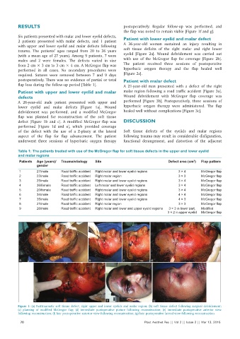
RESULTS                                             postoperatively.  Regular  follow‑up  was  performed,  and
                                                              the flap was noted to remain viable [Figure 1f and g].
          Six patients presented with malar and lower eyelid defects,   Patient with lower eyelid and malar defect
          2 patients presented with malar defects, and 1 patient   A  36‑year‑old woman  sustained an  injury  resulting  in
          with  upper and lower eyelid  and malar defects following
          trauma.  The patients’  ages  ranged from 20 to 36  years   soft  tissue  deficits  of the  right  malar and right  lower
          (with a mean age of 27 years). Among 9 patients, 7 were   eyelid  [Figure  2a]. Wound  debridement was carried  out
          males and 2 were females.  The defects varied in size   with  use of the McGregor flap for coverage  [Figure  2b].
          from 2 cm × 3 cm to 3 cm × 4 cm. A McGregor flap was   The patient  received three sessions  of postoperative
          performed in  all cases.  No  secondary  procedures  were   hyperbaric oxygen  therapy  and the  flap healed well
          required. Sutures were removed between  7 and 9  days   [Figure 2c].
          postoperatively. There was no evidence of partial or total   Patient with malar defect
          flap loss during the follow‑up period [Table 1].    A 21‑year‑old  man presented with a defect of the right
          Patient with upper and lower eyelid and malar       malar region following a road traffic accident [Figure 3a].
          defects                                             Wound debridement with McGregor flap coverage was
          A 26‑year‑old male  patient  presented  with  upper and   performed  [Figure  3b]. Postoperatively, three sessions  of
          lower eyelid and malar defects  [Figure  1a]. Wound   hyperbaric oxygen therapy were administered. The flap
          debridement was performed, and a modified McGregor   healed well without complications [Figure 3c].
          flap  was planned for reconstruction of the soft tissue
          defect  [Figure  1b and c]. A  modified McGregor flap  was   DISCUSSION
          performed  [Figure  1d and e], which provided coverage
          of the  defect with  the  use  of  a  Z‑plasty at  the  lateral   Soft tissue defects of the eyelids and malar regions
          aspect of  the  flap for  flap advancement.  The  patient   following trauma may result in considerable disfiguration,
          underwent three sessions  of hyperbaric oxygen  therapy   functional derangement,  and distortion  of the  adjacent

          Table 1: The patients treated with use of the McGregor flap for soft tissue defects in the upper and lower eyelid
          and malar regions
           Patients  Age (years)/  Trauma/etiology  Site                            Defect area (cm )  Flap pattern
                                                                                                 2
                   gender
           1       27/male     Road traffic accident  Right malar and lower eyelid regions  3 × 4    McGregor flap
           2       23/male     Road traffic accident  Right malar region                  3 × 3      McGregor flap
           3       25/male     Road traffic accident  Right malar and lower eyelid regions  3 × 4    McGregor flap
           4       36/female   Road traffic accident  Left malar and lower eyelid regions  3 × 4     McGregor flap
           5       20/female   Road traffic accident  Right malar and lower eyelid regions  3 × 4    McGregor flap
           6       34/male     Road traffic accident  Right malar and lower eyelid regions  4 × 4    McGregor flap
           7       35/male     Road traffic accident  Right malar and lower eyelid regions  4 × 3    McGregor flap
           8       21/male     Road traffic accident  Right malar region                  3 × 3      McGregor flap
           9       26/male     Road traffic accident  Right malar and lower and upper eyelid regions  3 × 3 in lower part,   Modified
                                                                                    3 × 2 in upper eyelid  McGregor flap


           a                                 b                c             d


                  e                                   f                      g
          Figure 1: (a) Posttraumatic soft tissue defect, right upper and lower eyelids and malar region; (b) soft tissue defect following surgical debridement;
          (c) planning of modified McGregor flap; (d) immediate  postoperative picture following reconstruction; (e) immediate  postoperative anterior view
          following reconstruction; (f) late postoperative anterior view following reconstruction; (g) late postoperative lateral view following reconstruction
            70                                                           Plast Aesthet Res || Vol 2 || Issue 2 || Mar 13, 2015