
Page 28 - Read Online
P. 28
Friedman et al. Plast Aesthet Res 2022;9:58 https://dx.doi.org/10.20517/2347-9264.2022.77 Page 7 of 11
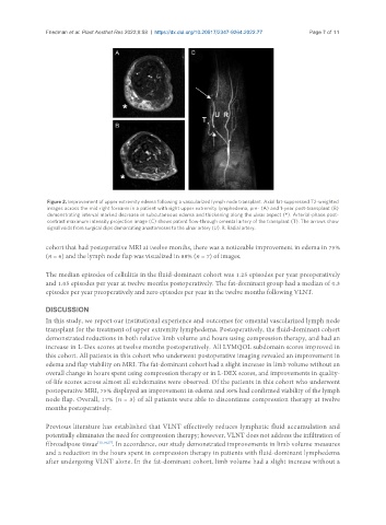
Figure 2. Improvement of upper extremity edema following a vascularized lymph node transplant. Axial fat-suppressed T2-weighted
images across the mid right forearm in a patient with right upper extremity lymphedema, pre- (A) and 1-year post-transplant (B)
demonstrating interval marked decrease in subcutaneous edema and thickening along the ulnar aspect (*). Arterial-phase post-
contrast maximum intensity projection image (C) shows patent flow-through omental artery of the transplant (T). The arrows show
signal voids from surgical clips demarcating anastomoses to the ulnar artery (U). R: Radial artery.
cohort that had postoperative MRI at twelve months, there was a noticeable improvement in edema in 75%
(n = 6) and the lymph node flap was visualized in 88% (n = 7) of images.
The median episodes of cellulitis in the fluid-dominant cohort was 1.25 episodes per year preoperatively
and 1.05 episodes per year at twelve months postoperatively. The fat-dominant group had a median of 0.3
episodes per year preoperatively and zero episodes per year in the twelve months following VLNT.
DISCUSSION
In this study, we report our institutional experience and outcomes for omental vascularized lymph node
transplant for the treatment of upper extremity lymphedema. Postoperatively, the fluid-dominant cohort
demonstrated reductions in both relative limb volume and hours using compression therapy, and had an
increase in L-Dex scores at twelve months postoperatively. All LYMQOL subdomain scores improved in
this cohort. All patients in this cohort who underwent postoperative imaging revealed an improvement in
edema and flap viability on MRI. The fat-dominant cohort had a slight increase in limb volume without an
overall change in hours spent using compression therapy or in L-DEX scores, and improvements in quality-
of-life scores across almost all subdomains were observed. Of the patients in this cohort who underwent
postoperative MRI, 75% displayed an improvement in edema and 88% had confirmed viability of the lymph
node flap. Overall, 17% (n = 3) of all patients were able to discontinue compression therapy at twelve
months postoperatively.
Previous literature has established that VLNT effectively reduces lymphatic fluid accumulation and
potentially eliminates the need for compression therapy; however, VLNT does not address the infiltration of
fibroadipose tissue [13,14,27] . In accordance, our study demonstrated improvements in limb volume measures
and a reduction in the hours spent in compression therapy in patients with fluid-dominant lymphedema
after undergoing VLNT alone. In the fat-dominant cohort, limb volume had a slight increase without a