
Page 25 - Read Online
P. 25
Page 4 of 11 Friedman et al. Plast Aesthet Res 2022;9:58 https://dx.doi.org/10.20517/2347-9264.2022.77
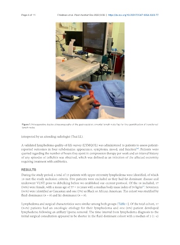
Figure 1. Intraoperative duplex ultrasonography of the gastroepiploic omental lymph node flap for the quantification of transferred
lymph nodes.
interpreted by an attending radiologist (Tsai LL).
A validated lymphedema quality-of-life survey (LYMQOL) was administered to patients to assess patient-
reported outcomes in four subdomains: appearance, symptoms, mood, and function . Patients were
[26]
queried regarding the number of hours they spent in compression therapy per week and an interval history
of any episodes of cellulitis was obtained, which was defined as an infection of the affected extremity
requiring treatment with antibiotics.
RESULTS
During the study period, a total of 23 patients with upper extremity lymphedema were identified, of which
18 met the study inclusion criteria. Five patients were excluded as they had fat-dominant disease and
underwent VLNT prior to debulking before we established our current protocol. Of the 18 included, 17
(94%) were female, with a mean age of 57 ± 10 years with a median body mass index of 30 kg/m . Seventeen
[2]
(94%) were identified as Caucasian and one (5%) as Black or African American. The cohort was stratified by
fluid-dominance (n = 9) and fat-dominance (n = 9).
Lymphedema and surgical characteristics were similar among both groups [Table 1]. Of the total cohort, 17
(94%) patients had an oncologic etiology for their lymphedema and one (6%) patient developed
lymphedema following an axillary lipoma removal. The time interval from lymphedema diagnosis to the
initial surgical consultation appeared to be shorter in the fluid-dominant cohort with a median of 2 (1-4)