
Page 34 - Read Online
P. 34
[5]
to be correlated with the risk of contralateral lymph node risk from 2.8 to 12.7). In the study of Kurita et al., patients
metastasis as well as with patient survival. We consider it with tumors showing radiological evidence of extension
important to analyze these factors. It is currently unclear crossing the midline were at a higher risk for CLNM (53.8%) than
whether CLNM are underestimated in OSCC patients at patients without an extension crossing the midline (10.3%).
initial presentation. Therefore, correct identification of risk
factors associated with CLNM is paramount to improve the In relation to the location of the primary tumor, a higher
clinical outcome of this patient group, especially because risk for CLNM in patients with tumors of the floor of the
ultrasound diagnostic imaging and computed tomography mouth and the anterior third of the tongue in detriment
scannings are not sensitive enough to sufficiently of the retromolar region or the lateral gum has been
[6]
detect occult disease. Prediction of tumors at high risk reported. Cross-drainage in the oral tongue and floor of
for contralateral involvement may determine a better mouth cancer is common, thereby placing both sides of
therapeutic management of the contralateral neck and may the neck at risk for nodal metastases, as reported in the
improve OSCC prognosis [Table 1]. study by Mukherji et al. Califano et al. found a higher
[22]
[10]
rate of contralateral involvement in the base of the
Tumor location tongue even in early tumors than in the body and the tip
One of the factors that has been speculated as a of the tongue and recommended prophylactic bilateral
determinant prognosticator for contralateral metastases neck dissection in all tongue base carcinomas. The data
[23]
is tumor location, although there is not a clear consensus of Olzowy et al. also showed that tumors of the base
about which location is of higher risk for cross-metastases. of tongue had a higher risk of contralateral metastases
than that of tumors of the tonsillar fossa. Moreover,
The importance of tumor midline involvement had been although not statistically significant, tumors of the soft
already exposed by Martin et al. Risk increased to 16% in palate and the pharyngeal walls also seemed to have a
[21]
cases with tumors crossing the midline by less than 1 cm and higher risk of CLNM. Capote-Moreno et al. observed a
[7]
reached 46% in those where the crossing was of more than higher tendency for contralateral metastases in tumors
[8]
1 cm. In the same way, Koo et al. also demonstrated that the located in the tongue base (31.4%) and the floor of the
rate of contralateral occult neck metastasis was significantly mouth (11%), with a lower frequency in the mobile tongue
higher in cases in which the primary lesion showed extension (7.2%) and the oropharynx (6.3%). However, in the study of
[5]
across the midline, compared with early-stage or Kurita et al., the incidence of CLNM was higher in cases
unilateral lesions. In a series including 513 consecutive of lower gum carcinoma (25%) than in those with mobile
cases, Kowalski et al. testified that the risks of CLNM were tongue carcinoma (15.4%). They suggested that the
[6]
significantly higher in cases of tumors extending to 1 cm or direction of tumor invasion is a more important factor for
less of the midline or crossing such medial margin (relative CLNM than the original tumor location in patients with
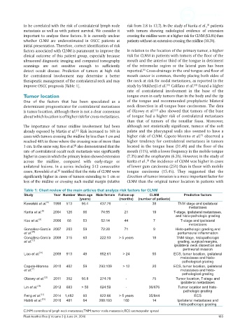
Table 1: Chart review of the main articles that analyze risk factors for CLNM
Study Year Number Mean age Male:female Follow-up CLNM Predictive factors
(years) (months) (number of patients)
Kowalski et al. [6] 1999 513 56.4 437:76 - 38 TNM stage and ipsilateral
metastases
Kurita et al. [5] 2004 126 66 74:55 21 19 T-stage, ipsilateral metastases,
and histo-pathologic grading
Koo et al. [8] 2006 66 53 52:14 44 7 T-stage and ipsilateral
metastases
González-García 2007 203 59 72:28 71 9 Histo-pathologic grading and
[20]
et al. peritumoural inflammation
González-García 2008 315 60 222:93 > 5 years 18 TNM stage, histopathologic
et al. [12] grading, surgical margins,
ipsilateral neck dissection and
perineural invasion
Liao et al. [31] 2009 913 49 852:61 > 24 55 ECS, tumor location, ipsilateral
metastases and histo-
pathological grading
Capote-Moreno 2010 402 59 293:109 > 12 20 ECS, tumor location, ipsilateral
et al. [7] metastases and histo-
pathological grading
[23]
Olzowy et al. 2011 352 56.8 274:78 - 75 Tumor location, T-stage and
ipsilateral metastases
Lin et al. [38] 2012 683 > 50 624:59 - 36/676 Tumor location and histo-
pathologic grading
[13]
Feng et al. 2014 1,482 60 822:66 > 5 years 35/844 ECS
Habib et al. [33] 2016 481 64 288:193 160 14 Ipsilateral metastases and
histo-pathologic grading
CLNM: contralateral lymph neck metastases; TNM: tumor node metastasis; ECS: extracapsular spread
Plast Aesthet Res || Volume 3 || June 24, 2016 183