
Page 34 - Read Online
P. 34
Page 6 of 12 Bizic et al. Plast Aesthet Res 2022;9:14 https://dx.doi.org/10.20517/2347-9264.2021.102
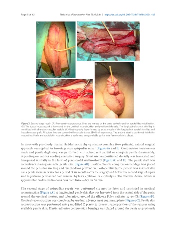
Figure 3. Second stage repair. (A) Preoperative appearance. Lines are marked on the penis ventrally and for scrotal flap mobilization.
(B) The buccal mucosa graft is harvested for the urethral reconstruction and positioned dorsally. The longitudinal scrotal skin flap is
mobilized with abundant vascular pedicle. (C) Urethroplasty is performed by anastomosis of the longitudinal scrotal skin flap and
buccal mucosa graft. All suture lines are covered with vascular tissue. (D) Final appearance. The urethral stent is positioned inside the
neourethra. Penile and scrotal skin reconstruction is performed using available genital skin. Penrose drain is placed.
In cases with previously treated bladder exstrophy epispadias complex (two patients), radical surgical
approach was applied for two-stage redo epispadias repair [Figure 4A and B]. Circumcision incision was
made and penile degloving was performed with subsequent partial or complete penile disassembly,
depending on entities needing corrective surgery. Short urethra positioned dorsally was transected and
transposed ventrally in the form of penoscrotal urethrostomy [Figure 4C and D]. The penile shaft was
reconstructed using available penile skin [Figure 4D]. Elastic-adhesive compression bandage was placed
around the penis for swelling and lymphedema prevention. Postoperatively, the patient was instructed to
use a penile vacuum device for a period of six months after the surgery and before the second stage of repair
and to perform permanent hair removal by laser epilation or electrolysis. The vacuum device, which is
approved for medical indications, was used twice a day for 30 min.
The second stage of epispadias repair was performed six months later and consisted in urethral
reconstruction [Figure 5A]. A longitudinal penile skin flap was harvested from the ventral side of the penis,
around the urethral meatus, and tubularized around the silicone Foley catheter 12-16 Ch [Figure 5B].
Urethral reconstruction was completed by urethral advancement and meatoplasty [Figure 5C]. Penile skin
reconstruction was performed using modified Z plasty to prevent superposition of the sutures using
available penile skin. Elastic-adhesive compression bandage was placed around the penis as previously