
Page 32 - Read Online
P. 32
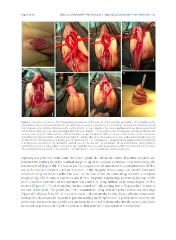
Page 4 of 12               Bizic et al. Plast Aesthet Res 2022;9:14  https://dx.doi.org/10.20517/2347-9264.2021.102


                Figure 1. Penopubic epispadias. (A) Preoperative appearance of the patient with penopubic epispadias. (B) Complete penile
                disassembly. Glans with neurovascular bundles. Both corpora cavernosa completely detached from the glans and mobilized urethral
                plate. Silicone urinary catheter placed inside the urethra. (C) Correction of dorsal curvature using grafting technique. Both corpora joined
                and derotated. Glans with two neurovascular bundles positioned dorsally. (D) The urethral plate is transposed ventrally and fixed to the
                corpora cavernosa. (E) Tubularization of the urethral plate over the silicone catheter. Glans is fixed to the corpora cavernosa.
                Glansplasty forming conical glans is finished. Gap between tubularized urethral plate and glans is visible and is approximately 2 cm long.
                (F) Mobilization of the preputial skin flap for urethral reconstruction. (G) Urethroplasty is completed using preputial island skin flap that
                is anastomosed proximally to the tubularized urethral plate and distally with the glandial part of the urethral plate. Tubularization is
                performed around the urinary catheter. Suture lines are covered with vascular subcutaneous tissue. (H) Outcome at the end of surgery.
                The urethral stent is positioned in the neourethra. The penile shaft is reconstructed using available penile skin.


               degloving was performed with caution to prevent penile skin devascularization. If urethra was short and
               presented the limiting factor for maximal straightening of the corpora cavernosa, it was transected at the
               subcoronal level [Figure 2B]. Artificial or pharmacological erection introduced by prostaglandin E1 (PDE1)
               was performed and recurrent curvature, present in the majority of redo cases, was noted . Curvature
                                                                                               [4]
               correction using bovine pericardium to cover the incision defects on tunica albuginea led to a complete
               straightening of both corpora cavernosa and allowed for penile lengthening, preventing shortage of the
               penis. Complete correction of the curvature was confirmed using artificial or pharmacological (PDE1)
               erection [Figure 2C]. The short urethra was transposed ventrally creating new a “hypospadiac” meatus at
               the base of the penis. The penile shaft was reconstructed using available penile and scrotal skin flaps
               [Figure 2D]. Silicone Foley Ch 12-16 catheter was introduced into the bladder. Elastic-adhesive compression
               bandage was placed around the penis to prevent swelling and lymphedema. In postoperative recovery, the
               patient was instructed to use a penile vacuum device for a period of six months after the surgery and before
               the second-stage repair and to perform permanent hair removal by laser epilation or electrolysis.