
Page 31 - Read Online
P. 31
Page 8 of 17 Toyoda et al. Plast Aesthet Res 2022;9:17 https://dx.doi.org/10.20517/2347-9264.2021.118
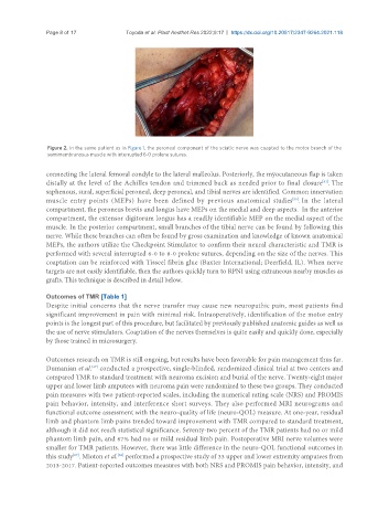
Figure 2. In the same patient as in Figure 1, the peroneal component of the sciatic nerve was coapted to the motor branch of the
semimembranosus muscle with interrupted 6-0 prolene sutures.
connecting the lateral femoral condyle to the lateral malleolus. Posteriorly, the myocutaneous flap is taken
distally at the level of the Achilles tendon and trimmed back as needed prior to final closure . The
[61]
saphenous, sural, superficial peroneal, deep peroneal, and tibial nerves are identified. Common innervation
muscle entry points (MEPs) have been defined by previous anatomical studies . In the lateral
[55]
compartment, the peroneus brevis and longus have MEPs on the medial and deep aspects. In the anterior
compartment, the extensor digitorum longus has a readily identifiable MEP on the medial aspect of the
muscle. In the posterior compartment, small branches of the tibial nerve can be found by following this
nerve. While these branches can often be found by gross examination and knowledge of known anatomical
MEPs, the authors utilize the Checkpoint Stimulator to confirm their neural characteristic and TMR is
performed with several interrupted 6-0 to 8-0 prolene sutures, depending on the size of the nerves. This
coaptation can be reinforced with Tisseel fibrin glue (Baxter International; Deerfield, IL). When nerve
targets are not easily identifiable, then the authors quickly turn to RPNI using extraneous nearby muscles as
grafts. This technique is described in detail below.
Outcomes of TMR [Table 1]
Despite initial concerns that the nerve transfer may cause new neuropathic pain, most patients find
significant improvement in pain with minimal risk. Intraoperatively, identification of the motor entry
points is the longest part of this procedure, but facilitated by previously published anatomic guides as well as
the use of nerve stimulators. Coaptation of the nerves themselves is quite easily and quickly done, especially
by those trained in microsurgery.
Outcomes research on TMR is still ongoing, but results have been favorable for pain management thus far.
[27]
Dumanian et al. conducted a prospective, single-blinded, randomized clinical trial at two centers and
compared TMR to standard treatment with neuroma excision and burial of the nerve. Twenty-eight major
upper and lower limb amputees with neuroma pain were randomized to these two groups. They conducted
pain measures with two patient-reported scales, including the numerical rating scale (NRS) and PROMIS
pain behavior, intensity, and interference short surveys. They also performed MRI neurograms and
functional outcome assessment with the neuro-quality of life (neuro-QOL) measure. At one-year, residual
limb and phantom limb pains trended toward improvement with TMR compared to standard treatment,
although it did not reach statistical significance. Seventy-two percent of the TMR patients had no or mild
phantom limb pain, and 67% had no or mild residual limb pain. Postoperative MRI nerve volumes were
smaller for TMR patients. However, there was little difference in the neuro-QOL functional outcomes in
this study . Mioton et al. performed a prospective study of 33 upper and lower extremity amputees from
[62]
[27]
2013-2017. Patient-reported outcomes measures with both NRS and PROMIS pain behavior, intensity, and