
Page 27 - Read Online
P. 27
Page 8 of 11 Hicks et al. Plast Aesthet Res 2022;9:2 https://dx.doi.org/10.20517/2347-9264.2021.65
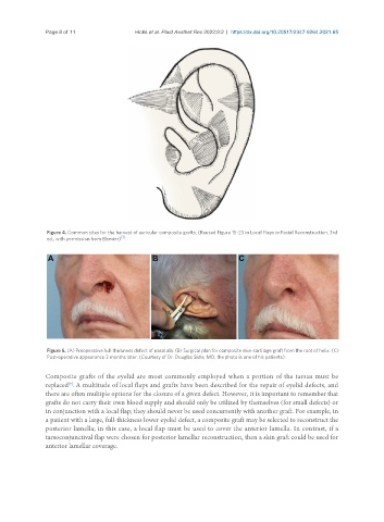
Figure 4. Common sites for the harvest of auricular composite grafts. (Reused Figure 15-23 in Local Flaps in Facial Reconstruction, 3rd
[1]
ed., with permission from Elsevier) .
Figure 5. (A) Preoperative full-thickness defect of nasal ala. (B) Surgical plan for composite skin-cartilage graft from the root of helix. (C)
Post-operative appearance 3 months later. (Courtesy of Dr. Douglas Sidle, MD, the photo is one of his patients).
Composite grafts of the eyelid are most commonly employed when a portion of the tarsus must be
replaced . A multitude of local flaps and grafts have been described for the repair of eyelid defects, and
[9]
there are often multiple options for the closure of a given defect. However, it is important to remember that
grafts do not carry their own blood supply and should only be utilized by themselves (for small defects) or
in conjunction with a local flap; they should never be used concurrently with another graft. For example, in
a patient with a large, full-thickness lower eyelid defect, a composite graft may be selected to reconstruct the
posterior lamella; in this case, a local flap must be used to cover the anterior lamella. In contrast, if a
tarsoconjunctival flap were chosen for posterior lamellar reconstruction, then a skin graft could be used for
anterior lamellar coverage.