
Page 24 - Read Online
P. 24
Hicks et al. Plast Aesthet Res 2022;9:2 https://dx.doi.org/10.20517/2347-9264.2021.65 Page 5 of 11
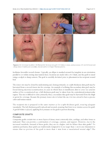
Figure 2. (A) Initial nasal tip defect. (B) Planned full-thickness skin graft. (C) Bolster in place, post-procedure. (D) and (E) frontal and
oblique views 4 months post-procedure. [Second author’s (J. Regan Thomas) patient].
facilitates favorable wound closure. Typically, an ellipse is marked around the template in an orientation
parallel to or within resting skin tension lines. Incisions are made with a #15 blade, and the graft is raised
using a scalpel or sharp scissors. The graft is carefully de-fatted prior to placement in the recipient wound
bed.
The donor site may be closed by undermining and closing primarily, or a split-thickness skin graft may be
harvested from a second donor site for coverage. An example of utilizing this secondary skin graft may be
found during microtia reconstruction; in cases in which there is insufficient skin to cover the anterior
surface of the reconstructed ear, a full-thickness graft may be taken from the contralateral postauricular
region. This site is difficult to close primarily; thus, a secondary skin graft may be harvested from the thigh
or groin for coverage. Because the primary donor site is behind the ear, it is less critical to have a precise
color and texture match.
The recipient site is prepared in the same manner as for a split-thickness graft, ensuring adequate
vascularity. The full-thickness graft is placed and secured, ensuring that there is no tension across the graft.
A gentle bolster is placed, applying firm pressure on the graft to prevent shearing.
COMPOSITE GRAFTS
Principles
Composite grafts contain two or more layers of tissue, most commonly skin, cartilage, and other tissue as
indicated. They can provide a combination of coverage, contour, and support. However, due to the
[5]
increased metabolic demand of these grafts, they are at a higher risk for failure than skin grafts . To
optimize perfusion, these grafts are traditionally designed to be no larger than 1 cm in any dimension to
ensure that no portion of the graft is more than 5 mm from a vascularized wound edge . The
[6]