
Page 16 - Read Online
P. 16
Sciegienka et al. Plast Aesthet Res 2022;9:1 https://dx.doi.org/10.20517/2347-9264.2021.76 Page 11 of 14
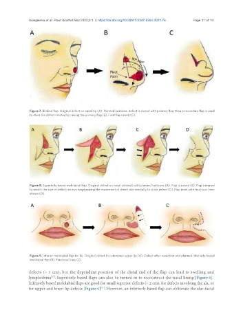
Figure 7. Bilobed flap. Original defect on nasal tip (A). Planned incisions; defect is closed with primary flap, then a secondary flap is used
to close the defect created by raising the primary flap (B). Final flap raised (C).
Figure 8. Superiorly based melolabial flap. Original defect on nasal sidewall with planned incisions (A). Flap is raised (B). Flap trimmed
to match the size of defect; arrows emphasizing the movement of cheek skin medially to close defect (C). Flap inset with final scar lines
shown (D).
Figure 9. Inferior melolabial flap for lip. Original defect in cutaneous upper lip (A). Defect after resection and planned inferiorly based
melolabial flap (B). Final scar lines (C).
defects (> 3 cm), but the dependent position of the distal end of the flap can lead to swelling and
[27]
lymphedema . Superiorly based flaps can also be turned in to reconstruct the nasal lining [Figure 8].
Inferiorly based melolabial flaps are good for small superior defects (< 2 cm), for defects involving the ala, or
[31]
for upper and lower lip defects [Figure 9] . However, an inferiorly based flap can obliterate the alar-facial