
Page 14 - Read Online
P. 14
Sciegienka et al. Plast Aesthet Res 2022;9:1 https://dx.doi.org/10.20517/2347-9264.2021.76 Page 9 of 14
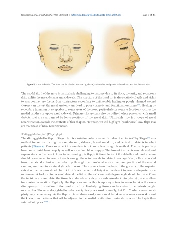
Figure 5. Nasal subunits. The nose can be divided into the tip, dorsal, columellar, and paired sidewall and alar lobules subunits.
The caudal third of the nose is particularly challenging to manage due to its thick, inelastic, and sebaceous
skin, unlike the nasal dorsum and sidewalls. The structure of the nasal tip is also relatively fragile and yields
to scar contracture forces. Scar contracture secondary to unfavorable healing or poorly planned wound
closure can distort the nasal anatomy and lead to poor cosmetic and functional outcomes . Healing by
[28]
secondary intention is acceptable in some areas of the nose, particularly in concave locations such as the
medial canthus or upper nasal sidewall. Primary closure may also be utilized when presented with small
defects that are surrounded by loose portions of the nasal skin. Ultimately, the full scope of nasal
reconstruction exceeds the contents of this chapter. However, we will highlight “workhorse” local flaps that
are mainstays of nasal reconstruction.
Sliding glabellar flap (Rieger flap)
The sliding glabellar flap or Rieger flap is a rotation-advancement flap described in 1967 by Rieger as a
[29]
method for reconstructing the nasal dorsum, sidewall, lateral nasal tip, and central tip defects in select
patients [Figure 6]. One can expect to close defects 2.5 cm or less using this method. The flap is partially
based on an axial blood supply as well as a random blood supply. The base of the flap is contralateral and
superolateral to the defect. Prior to performing this flap, soft tissue laxity of the glabella and nasal dorsum
should be evaluated to ensure there is enough tissue to provide full defect coverage. Next, a line is created
from the lateral extent of the defect up through the nasofacial sulcus, the nasal portion of the medial
canthus, and then to a natural glabellar crease. The distance from the base of the glabella to the superior
extent of the incision should be 1.5 to 2 times the vertical height of the defect to ensure adequate tissue
movement. A back cut to the contralateral medial canthus at about a 45-degree angle should be made. Once
the incisions are complete, the tissue is undermined widely in a submuscular (rhinoplasty) plane to allow
for maximum rotation. Typically, the flap is secured with a temporary suture to assess for skin thickness
discrepancy or distortion of the nasal structure. Underlying tissue can be excised to eliminate height
mismatches. The secondary glabellar defect can typically be closed primarily, but V to Y advancement or Z-
plasty may be necessary. As the flap is rotated downward, care should be taken to remove excess skin and
thickness from the tissue that will be adjacent to the medial canthus for maximal cosmesis. The flap is then
sutured into place [27,28] .