
Page 28 - Read Online
P. 28
a b
a b
c d c d
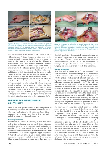
Figure 2: Technique of palmar cutaneous nerve relocation to pronator Figure 3: Technique of relocation of dorsal branch of ulnar nerve
quadratus. (a) Preoperative skin marking shows neuroma of the palmar into ulna. (a) End neuroma of dorsal branch of ulnar nerve; (b) nerve
cutaneous branch of the median nerve; (b) dissection of palmar dissected proximally; (c) nerve end can be placed in the ostium in distal
cutaneous nerve to origin from median nerve; (c) nerve relocated into ulna without tension; (d) epineurium is sutured to the periosteum to
muscle; (d) suture securing epineurium to epimysium prevent displacement of nerve end
tunnel is dissected in the muscle, and the nerve is buried than 50% conduction demonstrated intraoperatively across
without tension. A single absorbable suture between the the nerve. Opponents of neurolysis alone, however, warn
[33]
epineurium and epimysium holds the nerve in place. For of the risks of segmental revascularization and significant
placement into bone, a cortical hole is drilled obliquely to scar formation. This has led to the development of
[34]
a size slightly larger than the nerve diameter. The nerve techniques designed to prevent recurrent scarring such as
is relocated into this hole, and a single suture holds the wrapping the nerve in a variety of protective substances.
epineurium to the adjacent periosteum [Figure 3]. It is
important not to choose a site just distal to a joint where Nerve wrapping
[35]
[36]
mobilization is likely to put strain on the nerve. It is also First described by Masear et al., vein wrapping has
crucial to ensure there are no kinks or tension on the been reported as a successful technique in the management
nerve and that it does not angle acutely on entering the of both refractory cubital and carpal tunnel syndromes.
bone. Our primary preferences for nerve relocation are Initially, glutaraldehyde‑preserved allograft was used but
as follows: (1) superficial radial nerve to the undersurface has since been shown to cause increased scarring and
[37]
of brachioradialis or into radius; (2) palmar cutaneous adherence compared to autograft vein. Our unit has
branch of median nerve to pronator quadrates; (3) dorsal not had success with either technique and our limited
branch of ulnar nerve to pronator quadrates; (4) lateral experience of re‑exploration in these cases the nerve was
cutaneous nerve of the forearm to pronator quadrates; found to be tethered at both the proximal and distal sites
(5) digital nerves at or distal to distal interphalangeal joint on entry and exit of the vein graft. In general, we prefer to
to the proximal phalanx; and (6) digital nerves proximal use vascularized fascia to wrap nerves. The largest series of
[38]
the distal interphalangeal joint to the metacarpal shaft. vein wrapping to date has been reported by Kokkalis et al.
who performed the procedure on seventeen patients for
recalcitrant ulnar nerve compression at the elbow. Although
SURGERY FOR NEUROMAS‑IN‑ they reported a significant reduction in symptoms in most of
CONTINUITY the patients, pain was not abolished in any single case.
There is an even greater debate in the management of For neuromas involving the critical ulnar and median
in‑continuity neuromas. The treatment options fall into the nerves at the wrist or forearm, it has been our practice
following broad categories: (a) neurolysis alone, (b) nerve to use local fascial flaps to wrap the nerve, occasionally
wrapping, (c) neuroma resection and reconstruction, incorporating overlying skin and fat. Our unit has
and (d) neuroma resection and relocation. previously presented results of the analysis of 14 cases
of neurolysis and fascial wrapping for nerves‑in‑continuity
Neurolysis alone of the distal forearm or wrist. There was a complete
[39]
Preservation of functional sensitivity is vital for nerves resolution of pain in 8 of the 14 patients, and 2 more
such as the median and ulnar nerves. In these situations, patients had only mild pain at 6 month follow‑up. This
neurolysis alone or nerve wrapping usually maintains the technique was also used for the branches of the median
integrity of the intact axonal tissue. External neurolysis and ulnar nerves to the palm and digits in an effort to
theoretically restores movement, thereby, preventing further preserve distal sensation, using division and relocation
scar adherence to the nerve, a suggested trigger for pain. [31,32] as a secondary procedure in the case of failure. We
A study of neurolysis alone in upper trunk brachial plexus have found that there is little functional loss with the
neuroma‑in‑continuity revealed a functional improvement relocation of these nerves and have had greater success in
following neurolysis alone in cases where there was more abolishing pain with this technique.
168 Plast Aesthet Res || Vol 2 || Issue 4 || Jul 15, 2015