
Page 27 - Read Online
P. 27
Simple neuroma resection
Resection of the neuroma alone is the least successful
surgical method to treat neuromas of the hand and
forearm. However, laboratory studies have revealed
[16]
that the type of nerve transection can affect neuroma
formation. Neuromas developed more often after
electrocautery than simple scissors cut or suture
[17]
ligation and division. Decreased neuroma formation
and improved nerve regeneration have been noted
with oblique transection in comparison with transverse
sectioning for grafting. [18,19] It is suggested that the longer
fibers provide a growth pathway for the shorter ones with
oblique transection.
Containment
A number of methods of containment have been described
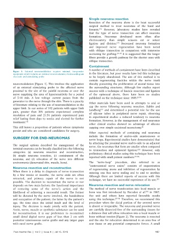
Figure 1: External neuromodulation requires minimal, inexpensive
equipment which includes an external neuromodulator, electrocardiogram in the literature, but poor results have led this technique
electrode and stimulating probe to be largely abandoned. The aim of this method is to
contain regenerating fascicles within the nerve trunk
neuromodulation [Figure 1]. This involves the application thereby preventing the proliferation of axonal tissue into
of an external stimulating probe to the affected nerve the surrounding structures. Although few studies report
proximal to the site of the painful neuroma or over the success with a technique of fascicle resection and ligation
nerve supplying the area of hypersensitivity for a period of the epineural sleeve, there have been no studies
of 5‑10 min. A low voltage current passes from the published on the technique since 1989. [8,20]
generator to the nerve through the skin. There is a paucity Other materials have been used in attempts to seal or
of literature relating to the use of neuromodulation in the cap the nerve following neuroma resection. Dahlin and
upper limb. In our series of 102 patients with upper limb Lundborg and determined a potential role for the use
[21]
pain, greater than 30% patients experienced complete of silicone tubes in peripheral nerve repair, observing
resolution of pain and 21.5% patients experienced pain in experimental studies a reduced tendency to neuroma
relief lasting from days to weeks and elected for further formation. However, in the management of end neuromas
treatment. [15]
other clinical studies showed no advantage of silicone
This still leaves a proportion of patients whose symptoms capping over simple excisional neurectomy. [8]
persist and who are considered candidates for surgery.
Other reported methods of containing end neuromas
include the formation of end‑to‑side anastomoses or
SURGERY FOR END‑NEUROMAS nerve loops. Experimental studies have demonstrated that
by attaching the proximal nerve end‑to‑side to an adjacent
The surgical options described for management of the nerve, the neuromas that form are smaller when compared
terminal neuroma can be broadly classified into the following to transection and epineural ligation. However, only
[22]
categories: (a) neuroma resection and reconstruction, preliminary clinical studies using this technique have been
(b) simple neuroma resection, (c) containment of the reported with small patient numbers. [23,24]
neuroma, and (d) relocation of the nerve into different
environments (denervated skin, muscle, bone). The “nerve‑loop” procedure, also referred to as
“centrocentral nerve union” consists of sequestration
Neuroma resection and reconstruction of regenerating axons and inhibition of regeneration by
When there is a delay in diagnosis of nerve transection suturing one free nerve ending end to end to another.
by a few weeks or months, the nerve ends are often Although there are limited reports of success with this
retracted, and primary repair of the nerve is not technique, we have no successful experience of its use. [25]
possible. The decision to reconstruct the nerve or nor
depends on two main factors: the functional importance Neuroma resection and nerve relocation
of restoring some of the nerve’s action and the The method of nerve translocation into local muscle or
likelihood of achieving a successful result. The former bone was first introduced by Herndon et al. in 1967.
[26]
will be dictated by the nerve involved, the handedness Our unit and others have reported favorable results
and occupation of the patient; the latter by the patient’s using this technique. [27‑30] Therefore, we recommend this
age, the time since the initial insult and the level of procedure when the distal portion of the severed nerve
injury. The decision is made jointly with the patient. is absent or irreparable. The neuroma and proximal nerve
Avein, nerve, muscle or synthetic substance can be used are carefully dissected free of the surrounding tissues for
for reconstruction. It is our preference to reconstruct a distance that will allow relocation into a local muscle or
small distal digital nerve gaps of less than 2 cm with bone without tension [Figure 2]. The neuroma is resected
posterior interosseous nerve grafts and any larger gaps and the site for relocation determined in an area free of
with sural nerve grafts. scar tissue or any potential compressive forces. A small
Plast Aesthet Res || Vol 2 || Issue 4 || Jul 15, 2015 167